. 2011 Aug 4;118(5):1294-304.
doi: 10.1182/blood-2011-01-330530. Epub 2011 May 11.

A B-cell subset uniquely responsive to innate stimuli accumulates in aged mice

Affiliations

A B-cell subset uniquely responsive to innate stimuli accumulates in aged mice

Yi Hao et al. Blood. .

Abstract

We have discovered a distinct mature B-cell subset that accumulates with age, which we have termed age-associated B cells. These cells comprise up to 30% of mature B cells by 22 months. Despite sharing some features with other mature B-cell subsets, they are refractory to BCR and CD40 stimulation. Instead, they respond to TLR9 or TLR7 stimulation and divide maximally on combined BCR and TLR ligation, leading to Ig production and preferential secretion of IL-10 and IL-4. Although similar to follicular B cells in both B-lymphocyte stimulator (BLyS) receptor expression and BLyS binding capacity, these cells do not rely on BLyS for survival. They are neither cycling nor the result of intrinsically altered B lymphopoiesis in aged BM, but instead appear to be generated from mature B cells that exhaustively expand during the individual's lifetime. Finally, they present Ag effectively and favor polarization to a TH17 profile. Together, these findings reveal that while the magnitude of the mature primary B-cell niche is maintained with age, it is increasingly occupied by cells refractory to BCR-driven activation yet responsive to innate receptor stimulation.


Figures

Figure 1
Figure 1
CD21/35CD23 B cells accumulate in aged mice. (A) Cells from spleen were first gated on CD19+ AA4.1 CD43 live single lymphocytes. Percentage of FO (CD21/35loCD23+), MZ (CD21/35hiCD23), and novel B-cell (CD21/35CD23, ABC) subsets are shown in female C57BL/6 mice at age 3, 12, and > 22 months. (B) Absolute numbers of ABCs in spleen from mice at age 3, 12, and > 22 months; n = 3-5 mice and similar results were observed in 5 independent experiments. The Student t test was used for statistical analysis; **P < .01. (C) Expression of 10 surface markers on ABCs (dark bold) compared with FO (dotted) and MZ (dashed) B cells from 22-month-old mice. Light gray peaks represent negative controls and the black peak represents the positive control for CD5 on B1 B cells.
Figure 2
Figure 2
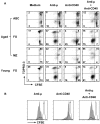
Impaired function of ABCs in vitro. (A) Flow cytometrically sorted splenic CD21/35CD23 (ABC) and FO B cells from aged or young mice were labeled with CFSE and then cultured with stimuli shown for 72 hours. Dead cells were stained by TO-PRO-3; CFSE dilution indicates proliferation. (B) Representative CFSE histograms for the same cultures shown in panel A show proliferation of ABCs (bold line) and FO (filled gray) B cells from aged mice. Similar results were found in 5 independent experiments.
Figure 3
Figure 3
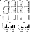
ABCs respond to TLR ligands in vitro. (A) Flow cytometrically sorted splenic ABCs from aged mice were labeled with CFSE then cultured with stimuli shown for 72 hours. Dead cells were stained by TO-PRO-3; CFSE dilution indicates proliferation. Similar results were found in 2 independent experiments. (B) CFSE histograms for the same cultures shown in panel A show proliferation of cells with (bold lines) or without (gray filled histograms) anti-μ. (C) TLR7 and TLR9 expression by qPCR in FO, MZ, and ABCs from young or aged mice. Fold change is relative to the level of each receptor observed in young FO (1.0 = the average level in FO B cells from young mice).
Figure 4
Figure 4
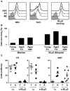
BLyS binding and BLyS requirement of ABCs. (A) Surface expression of BLyS receptors and BLyS binding capacity of ABCs (dark bold) compared with FO (dotted) and MZ (dashed) B cells from aged mice. (B) Flow cytometrically sorted splenic ABC and FO B cells from young and aged mice were cultured separately in the absence (medium only) or presence of BLyS. Living cells were TO-PRO-3 negative. (C) Effect of BLyS neutralization in vivo. Mice were treated with 100 μg of anti-BLyS or isotype control IgG1 IP on days 0 and 5. Numbers of FO, MZ and ABCs are shown at 21 days after the first injection. Mean with SEM are shown; each symbol represents one mouse. Similar results were observed from 3 independent experiments.
Figure 5
Figure 5
ABCs accumulate with age and can be generated from FO B cells on TLR stimulation in vitro. (A) Percentages of B-cell subsets in untreated young, aged, and autoreconstituted aged mice. The latter group of mice was sublethally irradiated (500R) on day 0 and allowed to reconstitute for 12 weeks. Gating strategy is the same as that described for Figure 1A. Similar results were observed in 3 independent experiments. (B) Cell-cycle analysis by DAPI for splenic ABC, FO, and BM pro-/pre- B cells from aged mice. (C) FO B cells from aged mice were sorted and CFSE-labeled, then cultured with stimuli shown for 3 days. Dead cells were excluded by DAPI staining. CD21/35 and CD23 expression on live cells is shown; CFSE dilution indicates cell division. CD19+ splenic B cells were analyzed on the same day as a reference for CD21/35 and CD23 expression (right-most plot).
Figure 6
Figure 6
FO B cells from both aged and young mice can give rise to ABCs after transfer into replete hosts. MACS-sorted CD23+ B cells (8 × 10⁶) from aged or young mice were labeled with CFSE, then injected separately into young congenic C57BL/6 hosts. Recipient mice were analyzed 1 month after transfer. (A) Percentages and numbers of donor cells 1 month after transfer. Top plots show live lymphocytes, CD19+B220+ gated, of donor (CD45.2+) and host (CD45.1+) phenotype. Absolute numbers of donor cells (bottom graph) were not significantly different (n.s.). Similar results were found in 5 experiments. (B) Surface-staining phenotype of CD45.2+ B cells before (left) and 1 month after transfer (right) shows ABCs (CD21/35CD23−) and MZ (CD21/35hiCD23) subsets of donor origin after 1 month. (C) CFSE dilution histograms of B cells from aged (filled gray) and young (bold line) donors recovered 1 month after transfer. (D) The dilution of CFSE in B cells from aged and young donors accompanied by surface expression of CD21/35, CD23, and IgM.
Figure 7
Figure 7
Cytokine expression and T-cell polarization on Ag presentation by ABCs. (A) Sorted ABCs and aged or young FO B cells were cultured with stimuli shown for 3 days. RNA was extracted and analyzed for expression of cytokines by qPCR. Fold change is relative to the expression level of each gene in standard total mouse RNA. (B) CFSE-labeled naive OTII CD4+ T cells (L-selectinhiCD44lo) were cultured with polarizing cytokines and Ab for 5 days, in the presence of young or aged FO or DC (CD11chi) that had been pulsed with ovalbumin323-339 peptide. Intracellular expression of cytokines and CFSE dilution were analyzed on gated CD4+ OTII T cells.

Comment in

  • Now you know your ABCs.
    Pillai S. Pillai S. Blood. 2011 Aug 4;118(5):1187-8. doi: 10.1182/blood-2011-06-355131. Blood. 2011. PMID: 21816835 No abstract available.

References

    1. Maue AC, Yager EJ, Swain SL, Woodland DL, Blackman MA, Haynes L. T-cell immunosenescence: lessons learned from mouse models of aging. Trends Immunol. 2009;30(7):301–305. - PMC - PubMed
    1. Cancro MP, Hao Y, Scholz JL, et al. B cells and aging: molecules and mechanisms. Trends Immunol. 2009;30(7):313–318. - PMC - PubMed
    1. Eaton SM, Burns EM, Kusser K, Randall TD, Haynes L. Age-related defects in CD4 T cell cognate helper function lead to reductions in humoral responses. J Exp Med. 2004;200(12):1613–1622. - PMC - PubMed
    1. Ahmed M, Lanzer KG, Yager EJ, Adams PS, Johnson LL, Blackman MA. Clonal expansions and loss of receptor diversity in the naive CD8 T cell repertoire of aged mice. J Immunol. 2009;182(2):784–792. - PMC - PubMed
    1. Zhang X, Fujii H, Kishimoto H, LeRoy E, Surh CD, Sprent J. Aging leads to disturbed homeostasis of memory phenotype CD8(+) cells. J Exp Med. 2002;195(3):283–293. - PMC - PubMed

Publication types

MeSH terms

Substances