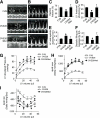
Improvement of cardiac functions by chronic metformin treatment is associated with enhanced cardiac autophagy in diabetic OVE26 mice
- PMID: 21562078
- PMCID: PMC3114402
- DOI: 10.2337/db10-0351
Improvement of cardiac functions by chronic metformin treatment is associated with enhanced cardiac autophagy in diabetic OVE26 mice
Abstract
Objective: Autophagy is a critical cellular system for removal of aggregated proteins and damaged organelles. Although dysregulated autophagy is implicated in the development of heart failure, the role of autophagy in the development of diabetic cardiomyopathy has not been studied. We investigated whether chronic activation of the AMP-activated protein kinase (AMPK) by metformin restores cardiac function and cardiomyocyte autophagy in OVE26 diabetic mice.
Research design and methods: OVE26 mice and cardiac-specific AMPK dominant negative transgenic (DN)-AMPK diabetic mice were treated with metformin or vehicle for 4 months, and cardiac autophagy, cardiac functions, and cardiomyocyte apoptosis were monitored.
Results: Compared with control mice, diabetic OVE26 mice exhibited a significant reduction of AMPK activity in parallel with reduced cardiomyocyte autophagy and cardiac dysfunction in vivo and in isolated hearts. Furthermore, diabetic OVE26 mouse hearts exhibited aggregation of chaotically distributed mitochondria between poorly organized myofibrils and increased polyubiquitinated protein and apoptosis. Inhibition of AMPK by overexpression of a cardiac-specific DN-AMPK gene reduced cardiomyocyte autophagy, exacerbated cardiac dysfunctions, and increased mortality in diabetic mice. Finally, chronic metformin therapy significantly enhanced autophagic activity and preserved cardiac functions in diabetic OVE26 mice but not in DN-AMPK diabetic mice.
Conclusions: Decreased AMPK activity and subsequent reduction in cardiac autophagy are important events in the development of diabetic cardiomyopathy. Chronic AMPK activation by metformin prevents cardiomyopathy by upregulating autophagy activity in diabetic OVE26 mice. Thus, stimulation of AMPK may represent a novel approach to treat diabetic cardiomyopathy.
Figures


Similar articles
-
AMP-activated protein kinase modulates cardiac autophagy in diabetic cardiomyopathy.Autophagy. 2011 Oct;7(10):1254-5. doi: 10.4161/auto.7.10.16740. Epub 2011 Oct 1. Autophagy. 2011. PMID: 21685727 Free PMC article.
-
Dissociation of Bcl-2-Beclin1 complex by activated AMPK enhances cardiac autophagy and protects against cardiomyocyte apoptosis in diabetes.Diabetes. 2013 Apr;62(4):1270-81. doi: 10.2337/db12-0533. Epub 2012 Dec 7. Diabetes. 2013. PMID: 23223177 Free PMC article.
-
Metformin Enhances Autophagy and Provides Cardioprotection in δ-Sarcoglycan Deficiency-Induced Dilated Cardiomyopathy.Circ Heart Fail. 2019 Apr;12(4):e005418. doi: 10.1161/CIRCHEARTFAILURE.118.005418. Circ Heart Fail. 2019. PMID: 30922066
-
Autophagy-dependent and -independent modulation of oxidative and organellar stress in the diabetic heart by glucose-lowering drugs.Cardiovasc Diabetol. 2020 May 13;19(1):62. doi: 10.1186/s12933-020-01041-4. Cardiovasc Diabetol. 2020. PMID: 32404204 Free PMC article. Review.
-
Targeting the energy guardian AMPK: another avenue for treating cardiomyopathy?Cell Mol Life Sci. 2017 Apr;74(8):1413-1429. doi: 10.1007/s00018-016-2407-7. Epub 2016 Nov 4. Cell Mol Life Sci. 2017. PMID: 27815596 Free PMC article. Review.
Cited by
-
Untangling autophagy measurements: all fluxed up.Circ Res. 2015 Jan 30;116(3):504-14. doi: 10.1161/CIRCRESAHA.116.303787. Circ Res. 2015. PMID: 25634973 Free PMC article. Review.
-
Metformin therapy in diabetes: the role of cardioprotection.Curr Atheroscler Rep. 2013 Apr;15(4):314. doi: 10.1007/s11883-013-0314-z. Curr Atheroscler Rep. 2013. PMID: 23423523 Review.
-
Cellular and molecular mechanisms of metformin: an overview.Clin Sci (Lond). 2012 Mar;122(6):253-70. doi: 10.1042/CS20110386. Clin Sci (Lond). 2012. PMID: 22117616 Free PMC article. Review.
-
Metformin alleviates bone loss in ovariectomized mice through inhibition of autophagy of osteoclast precursors mediated by E2F1.Cell Commun Signal. 2022 Oct 25;20(1):165. doi: 10.1186/s12964-022-00966-5. Cell Commun Signal. 2022. PMID: 36284303 Free PMC article.
-
Prenatal metformin exposure in mice programs the metabolic phenotype of the offspring during a high fat diet at adulthood.PLoS One. 2013;8(2):e56594. doi: 10.1371/journal.pone.0056594. Epub 2013 Feb 15. PLoS One. 2013. PMID: 23457588 Free PMC article.
References
-
- Levine B, Klionsky DJ. Development by self-digestion: molecular mechanisms and biological functions of autophagy. Dev Cell 2004;6:463–477 - PubMed
-
- Gustafsson AB, Gottlieb RA. Mechanisms of apoptosis in the heart. J Clin Immunol 2003;23:447–459 - PubMed
-
- Nakai A, Yamaguchi O, Takeda T, et al. The role of autophagy in cardiomyocytes in the basal state and in response to hemodynamic stress. Nat Med 2007;13:619–624 - PubMed
-
- UK Prospective Diabetes Study (UKPDS) Group Effect of intensive blood-glucose control with metformin on complications in overweight patients with type 2 diabetes (UKPDS 34). Lancet 1998;352:854–865 - PubMed
Publication types
MeSH terms
Substances
Grants and funding
- R01 HL089920/HL/NHLBI NIH HHS/United States
- R01 HL074399/HL/NHLBI NIH HHS/United States
- R01 HL079584/HL/NHLBI NIH HHS/United States
- HL-080499/HL/NHLBI NIH HHS/United States
- HL-074399/HL/NHLBI NIH HHS/United States
- R01 HL080499/HL/NHLBI NIH HHS/United States
- 1P20-RR-02421501/RR/NCRR NIH HHS/United States
- HL-096032/HL/NHLBI NIH HHS/United States
- HL-079584/HL/NHLBI NIH HHS/United States
- R01 HL110488/HL/NHLBI NIH HHS/United States
- R01 HL105157/HL/NHLBI NIH HHS/United States
- HL-089920/HL/NHLBI NIH HHS/United States
- R01 HL096032/HL/NHLBI NIH HHS/United States
LinkOut - more resources
Full Text Sources
Medical
Molecular Biology Databases

