Comparative Study
. 2011 Aug;106(2):630-40.
doi: 10.1152/jn.00015.2010. Epub 2011 May 11.

The magnitudes of hyperpolarization-activated and low-voltage-activated potassium currents co-vary in neurons of the ventral cochlear nucleus

Affiliations
Comparative Study

The magnitudes of hyperpolarization-activated and low-voltage-activated potassium currents co-vary in neurons of the ventral cochlear nucleus

Xiao-Jie Cao et al. J Neurophysiol. 2011 Aug.

Abstract

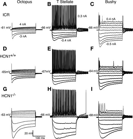
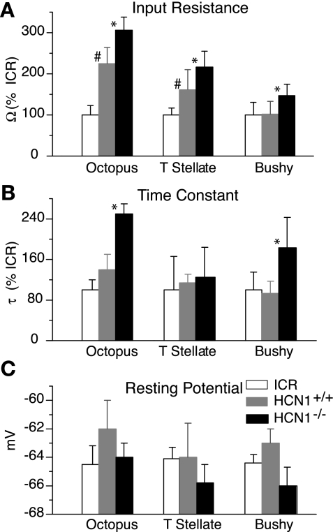
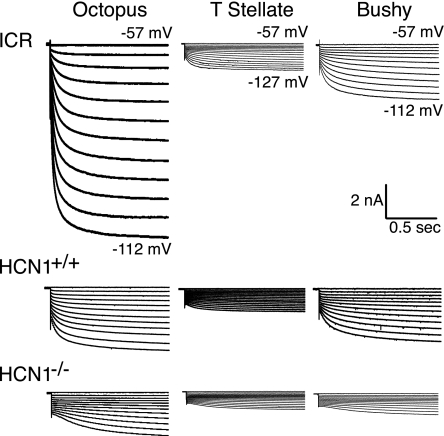
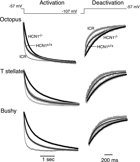
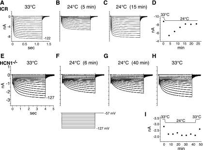
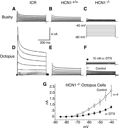
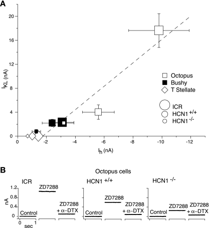
In the ventral cochlear nucleus (VCN), neurons have hyperpolarization-activated conductances, which in some cells are enormous, that contribute to the ability of neurons to convey acoustic information in the timing of their firing by decreasing the input resistance and speeding-up voltage changes. Comparisons of the electrophysiological properties of neurons in the VCN of mutant mice that lack the hyperpolarization-activated cyclic nucleotide-gated channel α subunit 1 (HCN1(-/-)) (Nolan et al. 2003) with wild-type controls (HCN1(+/+)) and with outbred ICR mice reveal that octopus, T stellate, and bushy cells maintain their electrophysiological distinctions in all strains. Hyperpolarization-activated (I(h)) currents were smaller and slower, input resistances were higher, and membrane time constants were longer in HCN1(-/-) than in HCN1(+/+) in octopus, bushy, and T stellate cells. There were significant differences in the average magnitudes of I(h), input resistances, and time constants between HCN1(+/+) and ICR mice, but the resting potentials did not differ between strains. I(h) is opposed by a low-voltage-activated potassium (I(KL)) current in bushy and octopus cells, whose magnitudes varied widely between neuronal types and between strains. The magnitudes of I(h) and I(KL) were correlated across neuronal types and across mouse strains. Furthermore, these currents balanced one another at the resting potential in individual cells. The magnitude of I(h) and I(KL) is linked in bushy and octopus cells and varies not only between HCN1(-/-) and HCN1(+/+) but also between "wild-type" strains of mice, raising the question to what extent the wild-type strains reflect normal mice.


Figures

Fig. 1.
Fig. 1.
Responses to current pulses reflect the intrinsic electrical properties of neurons. A–C: recordings from octopus, T stellate, and bushy cells in ICR mice illustrate the differences that distinguish them. Depolarizing current pulses caused octopus cells to fire transiently with a single action potential, bushy cells to fire transiently with several action potentials, and T stellate cells to fire tonically. Hyperpolarizing current pulses resulted in a polarization that sags back toward rest, reflecting the activation of hyperpolarization-activated (Ih) current. D–F: responses to current in neurons from hyperpolarization-activated cyclic nucleotide-gated 1 channel wild-type control (HCN1+/+) mice are generally similar to those in ICR mice, reflecting the characteristic differences among octopus, T stellate, and bushy cells. G–I: corresponding recordings from neurons in mice that lack the HCN1 α subunit (HCN1−/−) also show similar overall characteristics. Current pulses evoke larger polarizations in HCN1−/− mice than in the strains that contain HCN1, reflecting their higher input resistances. Amplitudes of current pulses, given in the upper panels, were matched in ICR, HCN1+/+, and mutant HCN1−/− neurons.
Fig. 2.
Fig. 2.
Comparison of characteristics of principal cells from ICR, HCN1+/+, and mutant HCN1−/− mice. A: input resistances (Ω) were significantly higher in HCN1−/− mutants than in HCN1+/+ in all 3 types of principal cells. The input resistances were also significantly higher in octopus and T stellate cells of HCN1+/+ than in ICR mice. B: the time constants (τ) were longer in octopus and bushy cells of HCN1−/− than in the HCN1+/+. Time constants were not significantly different in ICR and HCN1+/+ in any of the cell types. C: there were no significant differences in the resting potentials (mV) of neurons in the 3 strains of mice. Mean values ± SD are indicated by bars. *Statistically significant differences (P < 0.05) between mutant HCN1−/− mice and HCN1+/+, straddling the bars being compared. #Statistically significant differences (P < 0.05) between the HCN1+/+ and ICR mice.
Fig. 3.
Fig. 3.
Ih is smaller and slower in HCN1−/− mutant neurons than in HCN1+/+ or ICR neurons. Examples of recordings of Ih evoked by hyperpolarizing voltage steps from the holding potential of −57 mV in 5-mV increments to the levels indicated by numbers at the right of the traces. The steps evoked an instantaneous current that was proportional to the input conductance of the neurons; the inward current then increased gradually as Ih was activated. Ih was largest in octopus cells, intermediate in bushy cells, and smallest in T stellate cells of HCN1+/+ and ICR mice; in HCN1−/− mutant mice, Ih was similar in T stellate and bushy cells. Note that Ih activated more slowly in HCN1−/− neurons that lack HCN1 (bottom panels) than in HCN1+/+ or ICR neurons (middle and top panels, respectively). Recordings were made in the presence of 40 μM DNQX, 1 μM strychnine, 1 μM TTX, 0.25 mM Cd2+, and 50 nM α-DTX.
Fig. 4.
Fig. 4.
In all 3 groups of principal cells of the ventral cochlear nucleus, the kinetics of the activation and deactivation of Ih are slower in HCN1−/− than in HCN1+/+, and they are slower in HCN1+/+ than in ICR mice. Left: traces show responses to voltage steps from −57 mV to −107 mV in examples of each of the 3 cell types. Traces were fit with the sum of 2 exponential functions from the beginning of the activation of Ih to near the steady-state; panels illustrate only the early parts of those traces and the double exponential fit. Recordings from individual cells from the 3 strains of mice are superimposed for comparison. The heavy lines are double exponential fits to HCN1−/− cells, intermediate lines are fits to HCN1+/+, and the finest lines are fits to ICR mice. Right: traces show tail currents when the voltage was stepped back from −107 to −57 mV and were fit with single exponential functions. The heavy lines show fits to traces from mutant HCN1−/− cells, intermediate lines show the fits to HCN1+/+, and the finest lines show fits to traces from ICR mice.
Fig. 5.
Fig. 5.
Mouse strains differ in regulation of the magnitude of Ih in octopus cells. A–C: in an octopus cell from an ICR mouse, the temperature was shifted from 33°C to 24°C, and the cell's properties were assayed by stepping the voltage from −57 mV to a range of voltages between −57 and −122 mV. Five minutes after the temperature was reduced to 24°C, the same voltage steps evoked currents that were slower and smaller. Over the next 15 min, while the cell continued to be held at 24°C, the amplitude of Ih grew to near its original amplitude at the steady-state and remained slower than at 33°C. D: a plot of the amplitude of the steady-state current in response to a voltage step to −122 mV shows the time course with which the amplitude of the current returns to near the original value. E–H: a recording from an octopus cell of a HCN1−/− mouse that was subjected to a similar temperature change shows that the amplitude of Ih remained reduced for the 40 min after the temperature was reduced from 33°C to 24°C and that the change was largely reversed by elevating the temperature after that period. I: plot of the time course of the changes in Ih in the same HCN1−/− cell illustrated in E–H.
Fig. 6.
Fig. 6.
The magnitude of low-voltage-activated potassium (IKL) currents is reduced in neurons of HCN1−/− mice relative to HCN1+/+ in octopus and bushy cells. A: In a bushy cell from an ICR mouse, depolarizing voltage steps from −90 mV to −40 mV in 5-mV steps (shown in C) activated a voltage-sensitive outward current. B: in a bushy cell from a HCN1+/+ mouse, similar voltage steps evoked similar outward currents. C: In a bushy cell of a HCN1−/− mouse, outward currents evoked by similar voltage steps were smaller. Inset: voltage protocol applies to A–F. D: in an octopus cell from an ICR mouse, depolarizing voltage steps evoked large outward currents. E: similar voltage steps, applied to an octopus cell of a HCN1+/+ mouse, evoked smaller outward currents. F: in an octopus cell from a HCN1−/− mutant mouse, outward currents evoked by the same protocol were smaller still. Inset: more than one-half of the outward current was blocked by the application of 50 nM α-dendrotoxin (α-DTX). G: current/voltage relationship of average peak outward current in HCN1−/− octopus cells (n = 4) as a function of voltage under control conditions (o) and in the presence of 50 nM α-DTX (●). Recordings were made in the presence of 50 μM ZD7288, 1 μM TTX, 0.25 mM CdCl2, 40 μM DNQX, and 1 μM strychnine.
Fig. 7.
Fig. 7.
The magnitude of the IKL current is correlated with the magnitude of the opposing Ih inward currents across cell types and across strains of mice. A: the magnitude of Ih was assessed from the size of maximal inward currents evoked at the end of a 2-s voltage step from −57 mV to −122 mV in the presence of 40 μM DNQX, 1 μM strychnine, 1 μM TTX, 0.25 mM CdCl2, and 50 nM α-DTX. This current reflects the sum of Ih and a small leak current. The magnitude of IKL was measured in octopus and bushy cells as the peak outward current in response to voltage steps from −90 mV to −40 mV, measured in the presence of 40 μM DNQX, 1 μM strychnine, 1 μM TTX, 0.25 mM CdCl2, and 50 μM ZD7288. This measurement reflects the sum of IKL and a small leak current. The cell type is designated by the shapes of symbols, and the strain of mice is indicated by the relative size of symbols. Octopus cells of ICR mice have the largest Ih and also have the largest IKL, whereas bushy cells of HCN1−/− mutants have a small Ih and also a small IKL. A regression line, R = 0.95, was fit to measurements of Ih and IKL in octopus and bushy cells in all 3 stains of mice (dashed line). T stellate cells have no measurable IKL (Ferragamo and Oertel 2002); the values of Ih measured in T stellate cells in the 3 strains are shown with diamonds. B: in individual octopus cells, Ih and IKL are balanced at the resting potential. An octopus cell from each of the 3 strains of mice was initially held at the cell's resting potential. Resting potentials of the 3 octopus cells were: ICR −64 mV, HCN1+/+ −66 mV, and HCN1−/− −66 mV. Upon blocking the hyperpolarization-activated, mixed-cation conductance with 50 μM ZD7288, an outward holding current was observed. The further addition of 50 nM α-DTX in the continued presence of ZD7288 reduced the outward holding current to 0.1 nA. The similarity of the magnitudes of currents blocked by ZD7288 and α-DTX in individual cells indicates that in each cell, the inward Ih balanced the outward IKL at the holding potential.

References

    1. Adams JC. Projections from octopus cells of the posteroventral cochlear nucleus to the ventral nucleus of the lateral lemniscus in cat and human. Auditory Neurosci 3: 335–350, 1997
    1. Altomare C, Terragni B, Brioschi C, Milanesi R, Pagliuca C, Viscomi C, Moroni A, Baruscotti M, Altomare C, Terrangni B, Brioshi C, Milanesi R, Pegliuca C, Viscomi C, Moroni A, Baruscotti M, Baruscotti M, DiFrancesco D. Heteromeric HCN1-HCN4 channels: a comparison with native pacemaker channels from the rabbit sinoatrial node. J Physiol 549: 347–359, 2003 - PMC - PubMed
    1. Bal R, Baydas G, Naziroglu M. Electrophysiological properties of ventral cochlear nucleus neurons of the dog. Hear Res 256: 93–103, 2009 - PubMed
    1. Bal R, Oertel D. Hyperpolarization-activated, mixed-cation current (Ih) in octopus cells of the mammalian cochlear nucleus. J Neurophysiol 84: 806–817, 2000 - PubMed
    1. Bal R, Oertel D. Potassium currents in octopus cells of the mammalian cochlear nuclei. J Neurophysiol 86: 2299–2311, 2001 - PubMed

Publication types

MeSH terms

Substances

LinkOut - more resources