. 2011 May 31;108(22):9049-54.
doi: 10.1073/pnas.1018185108. Epub 2011 May 11.

Peptide surfactants for cell-free production of functional G protein-coupled receptors

Affiliations

Peptide surfactants for cell-free production of functional G protein-coupled receptors

Xiaoqiang Wang et al. Proc Natl Acad Sci U S A. .

Abstract

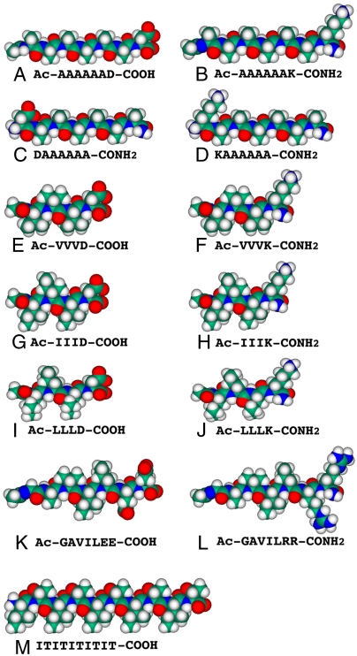
Two major bottlenecks in elucidating the structure and function of membrane proteins are the difficulty of producing large quantities of functional receptors, and stabilizing them for a sufficient period of time. Selecting the right surfactant is thus crucial. Here we report using peptide surfactants in commercial Escherichia coli cell-free systems to rapidly produce milligram quantities of soluble G protein-coupled receptors (GPCRs). These include the human formyl peptide receptor, human trace amine-associated receptor, and two olfactory receptors. The GPCRs expressed in the presence of the peptide surfactants were soluble and had α-helical secondary structures, suggesting that they were properly folded. Microscale thermophoresis measurements showed that one olfactory receptor expressed using peptide surfactants bound its known ligand heptanal (molecular weight 114.18). These short and simple peptide surfactants may be able to facilitate the rapid production of GPCRs, or even other membrane proteins, for structure and function studies.


Conflict of interest statement

The authors declare no conflict of interest.

Figures

Fig. 1.
Fig. 1.
Molecular models of the peptide surfactants and nonsurfactant peptide at pH 7.5. The peptide sequences are under each molecular model. Aspartic acid (D) and glutamic acid (E) are negatively charged whereas arginine (R) and lysine (K) are positively charged. The hydrophobic tails of the peptide surfactants consist of glycine (G), alanine (A), valine (V), isoleucine (I), and leucine (L). Each peptide surfactant is approximately 2–2.5 nm long, similar to biological phospholipids. Color code: teal, carbon; red, oxygen; blue, nitrogen; and white, hydrogen.
Fig. 2.
Fig. 2.
Western blotting detection of cell-free produced GPCRs. After the reactions, the samples were centrifuged. The supernatant containing the solubilized protein was removed, and the pellet was resuspended in an equivalent volume of buffer. The solubilized protein and resuspended pellets were detected by Western blotting. As controls, reactions with no peptide or the nonsurfactant peptide (IT)5 were performed. (A) Western blot of solubilized hFPR3 in the presence of different peptides. (B) Western blot of solubilized hOR17-210 produced in the presence of different peptides. Similar results were obtained with hTAAR5 and mOR103-15.
Fig. 3.
Fig. 3.
Comparison of the solubility of four GPCRs in 12 different peptide surfactants. Each GPCR was expressed in the presence of surfactant peptides using an E. Coli cell-free protein expression system. Upon completion, reactions were centrifuged to separate solubilized protein from aggregate. The soluble fraction was removed, and the pellet was resuspended in an equivalent volume of buffer. Solubilized and nonsolubilized protein fractions were assayed using a Western blot; relative band intensities were used to calculate the percentage of solubilized protein. As controls, reactions with no peptide or with the nonsurfactant peptide (IT)5 were performed. The presence of any peptide increased the fraction of solubilized protein. However, significant quantities of solubilized protein were only obtained in the presence of surfactant peptides.
Fig. 4.
Fig. 4.
The maximum expected yields of soluble receptors produced in the presence of peptide surfactants. (A) The maximum expected yields of solubilized monomer for each GPCR in the presence of each peptide or control condition. To determine the expected yields, solubilized protein and protein with a known concentration were assayed on a Western blot. The relative intensities of the known protein sample and the test samples were used to calculate the maximum protein yields. Surfactant peptides increased the yield of the solubilized monomeric form of the tested GPCRs. (B) The maximum yield of the monomeric form of each tested GPCR in a 10 mL reaction. Results from the most effective surfactant peptide are shown; it is compared to the maximum expected yield without peptide, or with a nonsurfactant peptide. The yield for each receptor varies with peptide surfactants used.
Fig. 5.
Fig. 5.
CD spectra of four GPCRs produced in the presence of peptide surfactants using a commercial E. coli cell-free system. (A) hFPR3 produced in Ac-VVVK-CONH2. (B) hTAAR5 produced in Ac-IIIK-CONH2. (C) mOR103-15 produced in Ac-VVVD-OH. (D) hOR17-210 produced in Ac-AAAAAAD-OH. These receptors all have characteristic α-helical spectra with valleys at 208 and 222 nm. Because GPCRs have 7-transmembrane α-helical domains, the CD spectra indicate that the receptors are properly folded.
Fig. 6.
Fig. 6.
Microscale thermophoresis ligand binding measurements of mOR103-15 produced in Ac-VVVD-OH. (A) Peptide-produced mOR103-15 has a sigmoidal shape, suggesting that the receptor binds to its ligand heptanal (MW 114.18). The measured EC50 is approximately 0.9 µM. (B) The boiled control showed fluctuations around a centerline, suggesting that binding observed with the normal receptor is not a result of nonspecific binding to the surfactant or denatured receptor. All curves were normalized to the fraction of bound receptor. Open circles show the mean measurements from three experiments; the lines through the points are the best-fit curves using the Hill equation.

References

    1. Wallin E, von Heijne G. Genome-wide analysis of integral membrane proteins from eubacterial, archaean, and eukaryotic organisms. Protein Sci. 1998;7:1029–1038. - PMC - PubMed
    1. Loll PJ. Membrane protein structural biology: The high throughput challenge. J Struct Biol. 2003;142:144–153. - PubMed
    1. Nilsson J, Persson B, von Heijne G. Comparative analysis of amino acid distributions in integral membrane proteins from 107 genomes. Proteins. 2005;60:606–616. - PubMed
    1. Vauthey S, Santoso S, Gong H, Watson N, Zhang S. Molecular self-assembly of surfactant-like peptides to form nanotubes and nanovesicles. Proc Natl Acad Sci USA. 2002;99:5355–5360. - PMC - PubMed
    1. Santoso S, Hwang W, Hartman H, Zhang S. Self-assembly of surfactant-like peptides with variable glycine tails to form nanotubes and nanovesicles. Nano Lett. 2002;2:687–691.

Publication types