. 2011 May 31;108(22):9238-43.
doi: 10.1073/pnas.1106200108. Epub 2011 May 11.

Helicobacter pylori cytotoxin-associated gene A (CagA) subverts the apoptosis-stimulating protein of p53 (ASPP2) tumor suppressor pathway of the host

Affiliations

Helicobacter pylori cytotoxin-associated gene A (CagA) subverts the apoptosis-stimulating protein of p53 (ASPP2) tumor suppressor pathway of the host

Ludovico Buti et al. Proc Natl Acad Sci U S A. .

Abstract

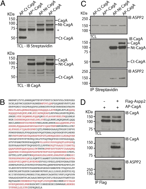
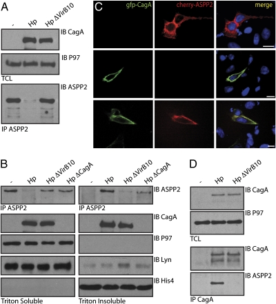
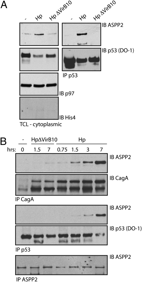
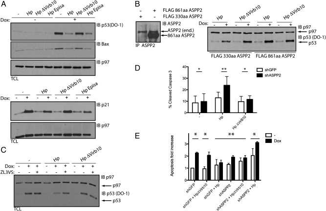
Type I strains of Helicobacter pylori (Hp) possess a pathogenicity island, cag, that encodes the effector protein cytotoxin-associated gene A (CagA) and a type four secretion system. After translocation into the host cell, CagA affects cell shape, increases cell motility, abrogates junctional activity, and promotes an epithelial to mesenchymal transition-like phenotype. Transgenic expression of CagA enhances gastrointestinal and intestinal carcinomas as well as myeloid and B-cell lymphomas in mice, but the mechanism of the induced cancer formation is not fully understood. Here, we show that CagA subverts the tumor suppressor function of apoptosis-stimulating protein of p53 (ASPP2). Delivery of CagA inside the host results in its association with ASPP2. After this interaction, ASPP2 recruits its natural target p53 and inhibits its apoptotic function. CagA leads to enhanced degradation of p53 and thereby, down-regulates its activity in an ASPP2-dependent manner. Finally, Hp-infected cells treated with the p53-activating drug Doxorubicin are more resistant to apoptosis than uninfected cells, an effect that requires ASPP2. The interaction between CagA and ASPP2 and the consequent degradation of p53 are examples of a bacterial protein that subverts the p53 tumor suppressor pathway in a manner similar to DNA tumor viruses. This finding may contribute to the understanding of the increased risk of gastric cancer in patients infected with Hp CagA+ strains.


Conflict of interest statement

The authors declare no conflict of interest.

Figures

Fig. 1.
Fig. 1.
CagA interacts with the tumor suppressor ASPP2. (A) Exogenous expression of BirA induces biotinylation of the AP-tagged CagA constructs. T-REx 293 stable transfectants were treated overnight with Dox to induce the expression of the indicated constructs together with the biotin ligase BirA. Total cell lysates were immunoblotted (IB) with streptavidin-HRP (Upper) and anti-CagA antibody (Lower). Full-length AP-CagA is partially processed into two fragments. Both full-length and N-terminal fragments are recognized by streptavidin-HRP. *Nonspecific band recognized by streptavidin-HRP. (B) AP-Nt CagA associates with endogenous ASPP2. Amino acid sequence of ASPP2. Peptides highlighted in bold were identified by LC-MS/MS in T-REx 293 expressing AP-Nt CagA and BirA. (C) Exogenously expressed CagA binds ASPP2 through its N-terminal domain. T-REx 293 stable transfectants were treated overnight with Dox to induce the expression of the indicated constructs together with BirA. Biotinylated proteins were retrieved by immunoprecipitation (IP) with streptavidin-conjugated beads. Total cell lysate (TCL) and IP were IB with the indicated antibodies. (D) HEK293T cells were cotransfected with FLAG-ASPP2 and AP-CagA. CagA was retrieved by IP with anti-CagA antibody, and immunoprecipitates were IB with the indicated antibodies.
Fig. 2.
Fig. 2.
CagA targets and relocalizes ASPP2 on Hp infection. Hp infection targets ASPP2. (A) AGS cells were infected for 6 h with the indicated Hp strains [multiplicity of infection (moi) = 1:100] or left uninfected and lysed in 1% Brij-containing buffer. TCLs were immunoblotted with the indicated antibodies. The ASPP2 input material is not shown because of the low expression level of the endogenous protein. ASPP2 was retrieved by IP with an anti-ASPP2 antibody, and the immunoprecipitates were IB with the indicated antibodies. (B) Hp infection induces relocalization of ASPP2 to a triton-resistant fraction. AGS cells were infected for 7 h with the indicated Hp strains (moi = 1:100) or left uninfected and lysed in Triton-X100 (T-X100) -containing buffer. The T-X100 insoluble fraction was then extracted in SDS buffer. Fractions were IB with the indicated antibodies or IP with anti-ASPP2 and IB with anti-ASPP2. P97 and Lyn serve as cytoplasmic markers, and HistoneH4 was a marker for nuclei. (C) Exogenously expressed CagA and ASPP2 colocalize near the plasma membrane. AGS cells cotransfected with FLAG-ASPP2 and Cherry-CagA are stained with anti-FLAG (green) and Hoechst (blue) (SI Materials and Methods and Fig. S2B). (Scale bar: 10 μm.) (D) Endogenous ASPP2 interacts with CagA during the course of in vivo Hp infection. AGS cells were infected for 7 h with the indicated Hp strains at an moi = 1:100 or left uninfected and lysed by sonication in Nonidet P-40–containing buffer. TCLs were immunoblotted with the indicated antibodies. The ASPP2 input material is not shown because of the low expression level of the endogenous protein. CagA was retrieved by IP with an anti-CagA antibody, and the immunoprecipitates were IB with the indicated antibodies.
Fig. 3.
Fig. 3.
ASPP2 associates with p53 on intracellular delivery of CagA. (A) Hp infection induces the association between endogenous p53 and ASPP2. AGS cells were infected with the indicated Hp strains (1:100 moi) for 7 h or left uninfected; then, they were sonicated in Nonidet P-40–containing buffer, and cytoplasmic p53 was IP. TCLs and immunoprecipitates were IB with the indicated antibodies, and p97 served as loading control as well as a cytoplasmic marker. His4 serves as a nuclear marker. (B) The CagA-ASPP2 interaction precedes the association between ASPP2 and p53. AGS cells were infected with the indicated Hp strains (moi = 1:100) or not infected, harvested at different time points, and processed as described in A. p53, CagA, and ASPP2 IP were IB with the indicated antibodies (SI Materials and Methods and Fig. S5A).
Fig. 4.
Fig. 4.
CagA inhibits p53 accumulation and transcriptional activity in an ASPP2-dependent manner. (A) CagA inhibits the accumulation and transcriptional activity of p53. HCT116p53+/+ cells were infected for 24 h with the indicated Hp strains (moi = 1:50) or left uninfected. Where indicated, cells were treated 5 h postinfection with 1 μg/mL Dox for 1.5 h. SDS-TCLs were IB with the indicated antibodies. p97 serves as loading control. We used HCT116 p53+/+ cells, because these can be infected with Hp (SI Materials and Methods and Fig. S6A) and their p53 pathway is well-characterized (41). (B) CagA inhibits accumulation of p53 in an ASPP2-dependent manner. HCT116 stably transfected with the indicated constructs were treated as indicated in A. To compare the level of endogenous ASPP2 with the overexpressed constructs, SDS lysates were IP and IB with ASPP2 antibody. This experiment is representative of three independent experiments. FLAG 330-aa ASPP2 construct is not shown, because the ASPP2 antibody does not recognize this short fragment (SI Materials and Methods and Fig. S4A). (C) CagA induces proteasomal degradation of p53. HCT116 cells were infected for 10 h with the indicated Hp strains (1:100 moi) or left uninfected. Where indicated, cells were treated 3 h postinfection with 1 μg/mL Dox for 1 h. Where indicated, ZL3VS was added for the remaining hours before harvesting the cells. TCLs were IB with the indicated antibodies. (D) ASPP2 inhibits apoptosis on Hp infection. Levels of cleaved Caspase-3 (CC3) assayed by flow cytometry in shGFP (white bars) or shASPP2 (construct #3; black bars) AGS cells that were infected for 24 h with the indicated Hp strains (1:50 moi) or left uninfected. Error bars ± SEM (n = 5). *P = ns; **P < 0.05. Significance was tested using two-way ANOVA Bonferroni multiple-comparison test. (E) Dox-induced apoptosis is inhibited by Hp in an ASPP2-dependent manner. Relative increase in CC3 in shGFP (white bars) or shASPP2 (construct #3; black bar) cells infected for 24 h with the indicated Hp strains (1:50 moi) or left uninfected and treated with 1 μg/mL Dox for 1 h. Error bars ± SEM (n = 3). *P < 0.05; **P = ns. Significance was tested using two-way ANOVA Bonferroni multiple-comparison test.

References

    1. Censini S, et al. cag, a pathogenicity island of Helicobacter pylori, encodes type I-specific and disease-associated virulence factors. Proc Natl Acad Sci USA. 1996;93:14648–14653. - PMC - PubMed
    1. Asahi M, et al. Helicobacter pylori CagA protein can be tyrosine phosphorylated in gastric epithelial cells. J Exp Med. 2000;191:593–602. - PMC - PubMed
    1. Odenbreit S, et al. Translocation of Helicobacter pylori CagA into gastric epithelial cells by type IV secretion. Science. 2000;287:1497–1500. - PubMed
    1. Stein M, Rappuoli R, Covacci A. Tyrosine phosphorylation of the Helicobacter pylori CagA antigen after cag-driven host cell translocation. Proc Natl Acad Sci USA. 2000;97:1263–1268. - PMC - PubMed
    1. Franco AT, et al. Regulation of gastric carcinogenesis by Helicobacter pylori virulence factors. Cancer Res. 2008;68:379–387. - PMC - PubMed

Publication types