. 2011 Jul 1;22(13):2175-84.
doi: 10.1091/mbc.E11-01-0031. Epub 2011 May 11.

Anaphase promoting complex-dependent degradation of transcriptional repressors Nrm1 and Yhp1 in Saccharomyces cerevisiae

Affiliations

Anaphase promoting complex-dependent degradation of transcriptional repressors Nrm1 and Yhp1 in Saccharomyces cerevisiae

Denis Ostapenko et al. Mol Biol Cell. .

Abstract

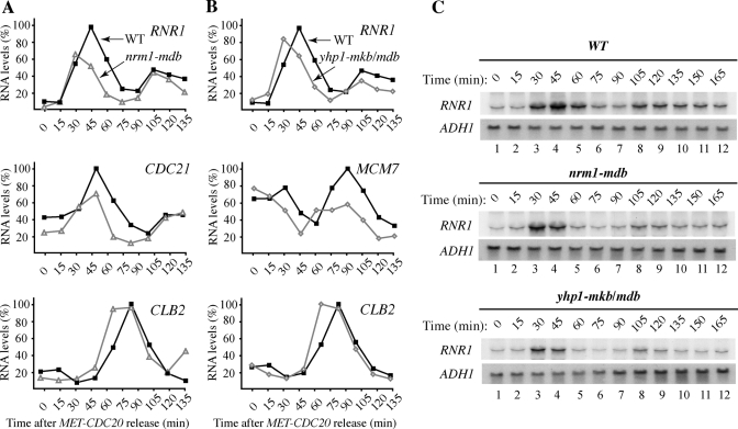
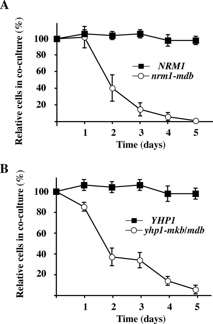
The anaphase-promoting complex/cyclosome (APC/C) is an essential ubiquitin ligase that targets cell cycle proteins for proteasome-mediated degradation in mitosis and G1. The APC regulates a number of cell cycle processes, including spindle assembly, mitotic exit, and cytokinesis, but the full range of its functions is still unknown. To better understand cellular pathways controlled by the APC, we performed a proteomic screen to identify additional APC substrates. We analyzed cell cycle-regulated proteins whose expression peaked during the period when other APC substrates were expressed. Subsequent analysis identified several proteins, including the transcriptional repressors Nrm1 and Yhp1, as authentic APC substrates. We found that APC(Cdh1) targeted Nrm1 and Yhp1 for degradation in early G1 through Destruction-box motifs and that the degradation of these repressors coincided with transcriptional activation of MBF and Mcm1 target genes, respectively. In addition, Nrm1 was stabilized by phosphorylation, most likely by the budding yeast cyclin-dependent protein kinase, Cdc28. We found that expression of stabilized forms of Nrm1 and Yhp1 resulted in reduced cell fitness, due at least in part to incomplete activation of G1-specific genes. Therefore, in addition to its known functions, APC-mediated targeting of Nrm1 and Yhp1 coordinates transcription of multiple genes in G1 with other cell cycle events.


Figures

FIGURE 1:
FIGURE 1:
Expression-based screening for potential APC substrates. (A) Flowchart showing the analysis of potential APC substrates. (B) The expression levels of endogenous Clb2-TAP and Clb1-TAP in extracts from asynchronous cells (lanes 1 and 4), cells arrested in G1 (lanes 2 and 5), and cells arrested in mitosis (lane 3 and 6) were compared by immunoblotting with anti-TAP antibodies. As a loading control, the membranes were reprobed with anti-PSTAIR antibodies to detect Cdc28 (lower panel). (C) Strains carrying selected TAP-tagged proteins were arrested in G1 and mitosis as above. Proteins that were at least threefold more abundant in benomyl-arrested cells than in G1-arrested cells (*) were considered potential candidates for further analyses.
FIGURE 2:
FIGURE 2:
Screening for proteins that are unstable in G1 but stable in mitosis. (A) Cells expressing the endogenously TAP-tagged proteins (as labeled) were arrested in G1 and treated with 500 μg/ml cycloheximide. Samples were withdrawn at the indicated times and processed for immunoblotting to detect TAP-tagged proteins. These proteins were stable and not considered further as potential APC substrates. (B) As in (A) but the stabilities of these proteins were analyzed both in G1 (as above) and in cells arrested in mitosis (M) by incubation with 20 μg/ml benomyl. Most of the proteins shown were unstable in G1 but stabilized in M phase, as expected for APC substrates. (C) cdh1-m11 targets Nrm1 and Yhp1 for unscheduled degradation. Cells carrying an empty vector or GALLp-cdh1-m11 were grown in the presence of raffinose and induced with 2% galactose for the indicated times. Cdh1-m11 lacks sites of inhibitory phosphorylation and is constitutively active. Samples were processed for immunoblotting to examine the endogenous levels of Clb2, Nrm1, and Yhp1. Cdc28 (*) was used as a loading control.
FIGURE 3:
FIGURE 3:
APC-dependent ubiquitination of Nrm1 and Yhp1. (A) Wild-type and cdc23–1 cells were arrested in G1 with α-factor and transferred to the nonpermissive temperature to inactivate Cdc23, a core subunit of the APC. cdc28–13 cdh1Δ cells were arrested in G1 by incubation at the nonpermissive temperature for cdc28–13. A putative D-box within Nrm1, 7RLPL, was mutated to generate Nrm1-mdb. The expression of NRM1 and nrm1-mdb were induced from GAL1p for 45 min followed by addition of 2% dextrose and 500 μg/ml cycloheximide to terminate protein synthesis. Samples were withdrawn at the indicated times and processed for immunoblotting with anti-TAP antibodies. Cdc28 (*) was used as a loading control. (B) Ubiquitination of Nrm1 in vitro. Nrm1 and Nrm1-mdb were synthesized in vitro in the presence of [35S]methionine and tested for ubiquitination in the absence (lanes 1 and 3) or presence (lanes 2 and 4) of purified APC and Cdh1. (C) Yeast two-hybrid interactions between Cdh1 and Yhp1. Cells expressing CDH1-ΔN200-DB or SNF1-DB (as a negative control) and YHP1-AD, HSL1-AD, or yhp1-mkb/mdb-AD were tested for growth on selective medium, which indicates interaction of the respective proteins. (D) Two potential degradation motifs, 329KEN-box and 340RKPL within Yhp1, were altered to generate Yhp1-mkb/mdb. Half-lives of Yhp1 and Yhp1-mkb/mdb in G1 were determined as in (A). Samples were withdrawn at the indicated times and processed for immunoblotting to detect Yhp1-TAP. (E) Ubiquitination of Yhp1 in vitro. 35S-labeled Yhp1 and Yhp1-mkb/mdb were tested for ubiquitination in the absence (lanes 1 and 3) or presence (lanes 2 and 4) of APC and Cdh1 as in (B).
FIGURE 4:
FIGURE 4:
Nrm1 and Yhp1 are degraded in G1 in a Cdh1-dependent manner. (A) cdc15–2 and cdc15–2 cdh1Δ cells carrying endogenous NRM1-TAP were synchronized in mitosis by incubation at 37°C for 3 h. Samples were withdrawn at the indicated times following release at 23°C and processed for immunoblotting to detect Nrm1-TAP. (B) MET-CDC20 cells carrying endogenous NRM1 or nrm1-mdb were synchronized in mitosis by incubation with 5 mM methionine for 2.5 h to deplete Cdc20. Cells were released from the arrest into methionine-free medium at time zero and samples were taken at the indicated times and processed for immunoblotting to detect Nrm1-TAP. Normalized levels of Nrm1 (solid squares) and Nrm1-mdb (open circles) were plotted below. The percentages of cells with anaphase spindles (shaded triangles) are indicated. (C) Yhp1 is degraded via APCCdh1 upon anaphase exit. cdc15–2 and cdc15–2 cdh1Δ cells carrying endogenous YHP1-TAP were synchronized and released from mitosis as in (A). Samples were withdrawn at the indicated times and processed for immunoblotting to detect Yhp1-TAP. (D) MET-CDC20 strains expressing endogenous YHP1 or yhp1-mkb/mdb were synchronized in mitosis as in (B). Samples were processed to detect Yhp1; Cdc28 was used as a loading control. Normalized levels of Yhp1 (solid squares) and Yhp1-mkb/mdb (open circles) are plotted. The percentages of cells with anaphase spindles (shaded triangles) are indicated.
FIGURE 5:
FIGURE 5:
Cdc28-mediated phosphorylation regulates Nrm1 stability. (A) GAL1p-NRM1 and nrm1-mdb were expressed in asynchronous cdc28-as cells in the presence of 2% galactose for 45 min followed by the addition of dimethyl sulfoxide (DMSO) (lanes 1–5) or 1 μM 1NM-PP1 (to inhibit Cdc28-as; lanes 6–10) for the last 10 min of galactose induction. The degradation of Nrm1 and Nrm1-mdb were examined following the addition of 2% dextrose and 500 μg/ml cycloheximide as in Figure 3A. (B) Four potential Cdc28 phosphorylation sites within Nrm1 were mutated to generate Nrm1–4TA. GALp-NRM1 and NRM1-4TA were expressed in asynchronous cells and Nrm1 and Nrm1-4TA degradation were examined as in (A). (C) The four potential Cdc28 phosphorylation sites were mutated to aspartic acid to generate Nrm1-4TD and the stabilities of Nrm1 and Nrm1-4TD in G1-arrested cells was assessed as in (B).
FIGURE 6:
FIGURE 6:
Nrm1 and Yhp1 degradation are required for proper G1 transcription. (A, B) Wild-type, nrm1-mdb, and yhp1-mkb/mdb strains were synchronized in M phase by CDC20 depletion, as in Figure 4B. Samples were withdrawn every 15 min after release and processed for quantitative reverse transcriptase PCR (qRT PCR) analysis with primers corresponding to RNR1, CDC21, MCM7, and CLB2. The relative level of each transcript was normalized to ACT1 mRNA in the same cells. (C) Northern blot analysis using 32P-labeled probes corresponding to RNR1 and ADH1 (for normalization).
FIGURE 7:
FIGURE 7:
Stabilization of Nrm1 and Yhp1 reduces cell fitness. (A) A genetically marked wild-type control strain was grown in coculture with a differentially marked NRM1 or nrm1-mdb test strain for 5 d with dilution of the cocultures once per day. The cultures were tested daily for the presence of TRP1 and LEU2 auxotrophs by plating on selective medium. The markers of the wild-type control strain and the experimental NRM1 and nrm1-mdb strains were swapped, the coculture experiment was repeated, and the results were averaged as described in Materials and Methods. (B) As in (A), but using YHP1 and yhp1-mkb/mdb test strains.

Similar articles

Cited by

References

    1. Belle A, Tanay A, Bitincka L, Shamir R, O’Shea EK. Quantification of protein half-lives in the budding yeast proteome. Proc Natl Acad Sci USA. 2006;103:13004–13009. - PMC - PubMed
    1. Benanti JA, Matyskiela ME, Morgan DO, Toczyski DP. Functionally distinct isoforms of Cik1 are differentially regulated by APC/C-mediated proteolysis. Mol Cell. 2009;33:581–590. - PMC - PubMed
    1. Biggs JR, Peterson LF, Zhang Y, Kraft AS, Zhang DE. AML1/RUNX1 phosphorylation by cyclin-dependent kinases regulates the degradation of AML1/RUNX1 by the anaphase-promoting complex. Mol Cell Biol. 2006;26:7420–7429. - PMC - PubMed
    1. Bishop AC, et al. A chemical switch for inhibitor-sensitive alleles of any protein kinase. Nature. 2000;407:395–401. - PubMed
    1. Burton JL, Solomon MJ. Hsl1p, a Swe1p inhibitor, is degraded via the anaphase-promoting complex. Mol Cell Biol. 2000;20:4614–4625. - PMC - PubMed

Publication types

MeSH terms

Substances