. 2011 Jul 1;22(13):2165-74.
doi: 10.1091/mbc.E10-11-0912. Epub 2011 May 11.

Transcriptional activation of histone H4 by C/EBPβ during the mitotic clonal expansion of 3T3-L1 adipocyte differentiation

Affiliations

Transcriptional activation of histone H4 by C/EBPβ during the mitotic clonal expansion of 3T3-L1 adipocyte differentiation

You-You Zhang et al. Mol Biol Cell. .

Abstract

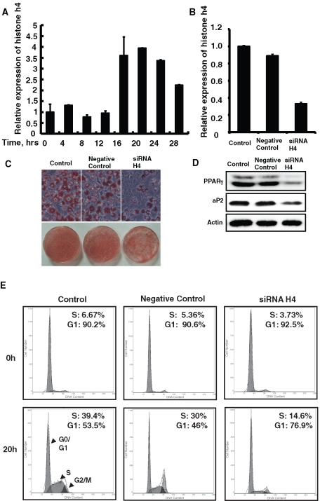
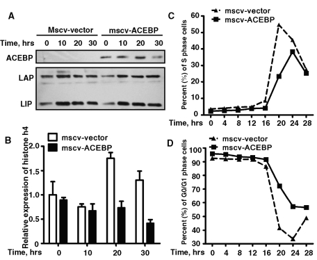
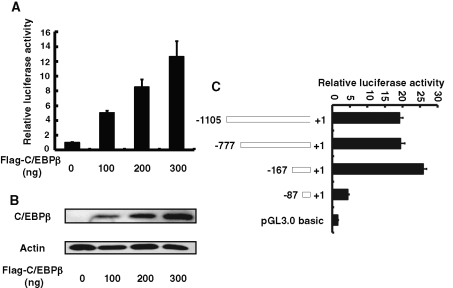
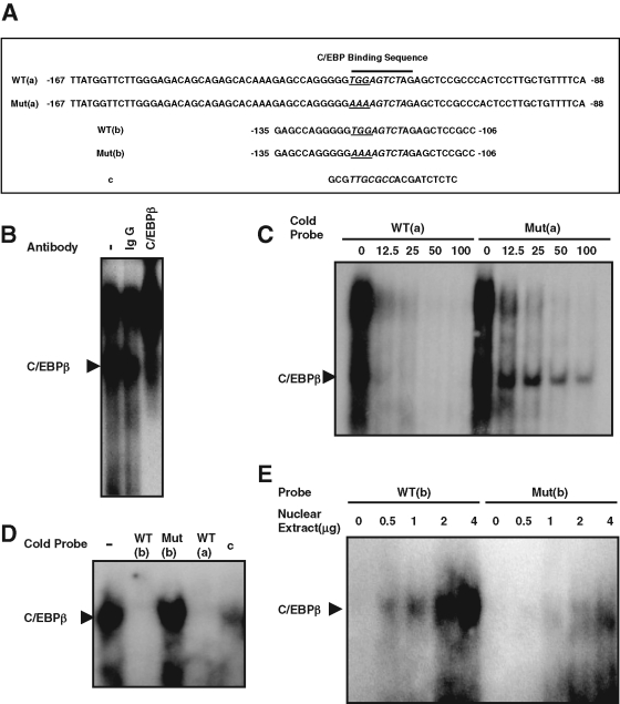
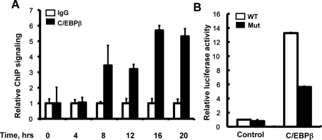
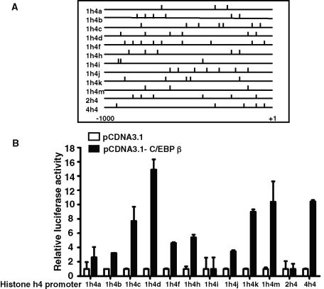
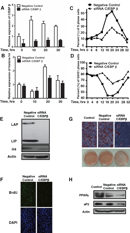
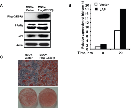
CCAAT enhancer binding protein β (C/EBPβ) is required for both mitotic clonal expansion (MCE) and terminal differentiation during the 3T3-L1 adipocyte differentiation program. Whereas the mechanism of C/EBPβ during terminal differentiation is well understood, the mechanism of C/EBPβ in MCE is not. We provide evidence that histone H4, the most conserved cell cycle-related histone, the change of which is strictly correlated with DNA content change during the cell cycle, is transcriptionally activated by C/EBPβ during MCE. Expression of histone H4 is increased at 16 h after induction when 3T3-L1 preadipocytes synchronously reenter S phase, which is correlated with the sequential phosphorylation and activation of C/EBPβ, and expression was partially suppressed when A-C/EBP (dominant negative for C/EBP protein) was overexpressed. One C/EBP-binding site was identified in one of the histone H4 gene promoters (hist4h4), confirmed by both electrophoretic mobility shift assay and chromatin immunoprecipitation assay. C/EBP-binding sites were also found in 9 of 11 other histone H4 promoters, which can also be transactivated by C/EBPβ. Knockdown of C/EBPβ by stealth small interfering RNA partially decreased H4 gene expression and arrested cells in G1 phase as indicated by bromodeoxyuridine incorporation and fluorescence-activated cell sorting analysis of DNA content. This study provides new insights into why C/EBPβ is required for MCE during 3T3-L1 adipocyte differentiation and why C/EBPβ plays important roles in the proliferation of other cell types.


Figures

FIGURE 1:
FIGURE 1:
The induction of histone H4 expression was required for mitotic clonal expansion and terminal differentiation of 3T3-L1 preadipocytes. (A) Postconfluent 3T3-L1 preadipocytes were induced to differentiate as described, total RNA was isolated at different time points, and quantitative real-time RT-PCR was performed to detect histone H4 expression. (B–E) 3T3-L1 preadipocytes were transfected with histone H4 siRNA, and 24 h after reaching confluence, cells were induced to differentiate as described. Total RNA was isolated at 20 h after induction, the expression of total H4 was detected by real-time RT-PCR, and 18S rRNA was used as loading control (B). After 8 d of induction, fat droplet accumulation was detected by oil red O staining and photographed (C), and the expression of PPAR γ and 422/aP2 was detected by Western blotting; actin was used as loading control (D). DNA content was analyzed by fluorescence-activated cell sorting, and the percentage of cells in S phase or G0/G1 phase was calculated at 0- and 20-h time points (E).
FIGURE 2:
FIGURE 2:
Overexpression of A-C/EBP partially suppressed the induction of histone H4 expression during MCE of 3T3-L1 adipocyte differentiation. (A, B) 3T3-L1 preadipocytes were infected with retrovirus expression flag–tagged A-C/EBP or with empty vector, cells were induced with differentiation media 2 d after reaching confluence, and the expression of A-C/EBP and C/EBPβ was detected by Western blotting (A); the effect of A-C/EBP on the expression of histone H4 was detected by quantitative real time RT-PCR as indicated in B. (C, D) DNA content was analyzed by fluorescence-activated cell sorting, and the percentage of cells in S phase or G0/G1 phase was analyzed and plotted.
FIGURE 3:
FIGURE 3:
The C/EBP response element was localized between −167 and −87 of the hist4h4 promoter. (A) and (B) 3T3-L1 preadipocytes were transiently transfected with hist4h4 promoter reporter construct, along with different amounts of C/EBPβ expression vector, and pRL-TK plasmid was used as internal control; cell extracts were prepared, and luciferase activities were measured and normalized to Renilla activity. (C) 3T3-L1 preadipocytes were transiently transfected with reporter constructs having different truncated hist4h4 promoter, together with C/EBPβ expression vector (300 ng). pRL-TK plasmid was used as internal control, and 48 h later, cell extracts were prepared and luciferase activity was measured and normalized to Renilla activity. Data are presented as means ± SD of at least three independent experiments.
FIGURE 4:
FIGURE 4:
One specific C/EBP-binding site was identified in the hist4h4 promoter. (A) The sequence of five oligonucleotides was presented for following EMSA. Postconfluent 3T3-L1 preadipocytes were induced to differentiate, and nuclear extract was prepared at 20 h. (B) EMSA was performed with probe WT(a); the lower band in lane 1 can be supershifted with specific C/EBPβ antibody in lane 3 but not by nonspecific IgG in lane 2. (C) Both bands (including the lower C/EBPβ band and the upper nonspecific band) were with equal efficiently competed away by cold oligonucleotide WT(a), whereas the lower C/EBPβ band was much more difficult to compete away than the upper nonspecific band by cold mutant oligonucleotide Mut(a), in which the C/EBP-binding site was mutated. (D) WT(b) was used as probe, and in lanes 2–5, 100-fold of cold probe WT(b), Mut(b), WT(a), and c were used for competition. (E) WT(b) and Mut(b) were used as probes, and EMSA was performed with 0–4 μg of nuclear extract EMSA.
FIGURE 5:
FIGURE 5:
Binding of C/EBPβ to the hist4h4 promoter was confirmed by ChIP. (A) Postconfluent 3T3-L1 preadipocytes were induced to differentiate as described, ChIP-qPCR was performed at different time points after induction with anti-C/EBPβ antibody, and nonspecific IgG was used as negative control. Data are normalized to the IgG controls at each time point. An area of the insulin gene served as a negative control. (B) 3T3-L1 preadipocytes were transiently transfected with wt-1105 pGL3.0-hist4h4 promoter plasmid (open bar) and its mutant plasmid (closed bar) in which the C/EBP-binding site was mutated from TTG to AAA, together with pCDNA3.1-C/EBPβ plasmid. Forty-eight hours later, cell extracts were prepared, and luciferase activity was measured and normalized to Renilla activity. Data are presented as means ± SD of at least three independent experiments.
FIGURE 6:
FIGURE 6:
Ten of 12 histone H4 promoters could be transactivated by C/EBPβ. (A) Analysis of the potential C/EBP-binding sites and their relative positions in an ∼1000–base pair length of all 12 histone H4 promoters, including hist4h4. (B) 3T3-L1 preadipocytes were transfected with pGL3.0-H4 promoter plasmids and pRL-TK plasmid, together with C/EBPβ expression plasmid (black bars) or empty vector (open bars). Forty-eight hours later, cell extracts were prepared, and luciferase activity was measured and normalized to Renilla activity. Data are presented as means ± SD of at least three independent experiments.
FIGURE 7:
FIGURE 7:
Knockdown of the expression of C/EBPβ with siRNA partially blocked the induction of histone H4, MCE, and adipocyte differentiation of 3T3-L1 preadipocytes. 3T3-L1 preadipocytes were transfected with C/EBPβ siRNA; 24 h after reaching confluence, cells were induced to differentiate as described. (A, B) Total RNA was isolated at various time points after induction; the expression of C/EBPβ and histone H4 was detected by real-time RT-PCR, with 18S rRNA used as loading control. (C, D) At different time points the DNA content was analyzed by fluorescence-activated cell sorting, and the percentages of cells in S phase or G0/G1 phase were calculated and plotted. (E) The expression of C/EBPβ and histone H4 was detected by Western blotting. (F) At 18 h after induction, a BrdU labeling experiment was performed and visualized with fluorescence microscopy. (G, H) After 8 d of induction, fat droplet accumulation was detected by oil red O staining, and the expression of PPAR γ and 422/aP2 was detected by Western blotting. Actin was used as loading control.
FIGURE 8:
FIGURE 8:
Overexpression of C/EBPβ promoted histone H4 expression and accelerated 3T3-L1 adipocyte differentiation. 3T3-L1 preadipocytes were infected with retrovirus expression flag–tagged C/EBPβ or with empty vector; cells were induced with differentiation media 2 d after reaching confluence, and the expression of C/EBPβ was detected by Western blotting (A); the effect of C/EBPβ on the expression of histone H4 was detected by quantitative real-time RT-PCR (B). Eight days after induction, fat droplet accumulation was detected by oil red O staining (C), and the expression of PPAR γ and 422/aP2 was detected by Western blotting; actin was used as loading control (A).

Similar articles

Cited by

References

    1. Aziz F, van Wijnen AJ, Stein JL, Stein GS. HiNF-D (CDP-cut/CDC2/cyclin A/pRB-complex) influences the timing of IRF-2-dependent cell cycle activation of human histone H4 gene transcription at the G1/S phase transition. J Cell Physiol. 1998a;177:453–464. - PubMed
    1. Aziz F, van Wijnen AJ, Vaughan PS, Wu SJ, Shakoori AR, Lian JB, Soprano KJ, Stein JL, Stein GS. The integrated activities of IRF-2 (HiNF-M), CDP/cut (HiNF-D) and H4TF-2 (HiNF-P) regulate transcription of a cell cycle controlled human histone H4 gene: mechanistic differences between distinct H4 genes. Mol Biol Rep. 1998b;25:1–12. - PubMed
    1. Birnbaum MJ, et al. Functional role for Sp1 in the transcriptional amplification of a cell cycle regulated histone H4 gene. Biochemistry. 1995;34:7648–7658. - PubMed
    1. Borun TW, Pearson D, Paik WK. Studies of histone methylation during HELA S-3 cell cycle. J Biol Chem. 1972;247:4288. - PubMed
    1. Buck M, Poli V, Van Der Geer P, Chojkier M, Hunter T. Phosphorylation of rat serine 105 or mouse threonine 217 in C/EBP beta is required for hepatocyte proliferation induced by TGF alpha. Mol Cell. 1999;4:1087–1092. - PubMed

Publication types

MeSH terms

Substances

LinkOut - more resources