. 2011 Sep;80(5):493-503.
doi: 10.1038/ki.2011.125. Epub 2011 May 11.

Acute loss of renal function attenuates slow leukocyte rolling and transmigration by interfering with intracellular signaling

Affiliations

Acute loss of renal function attenuates slow leukocyte rolling and transmigration by interfering with intracellular signaling

Jan Rossaint et al. Kidney Int. 2011 Sep.

Abstract

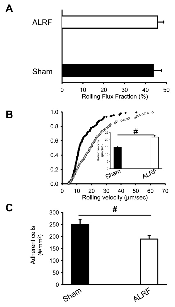
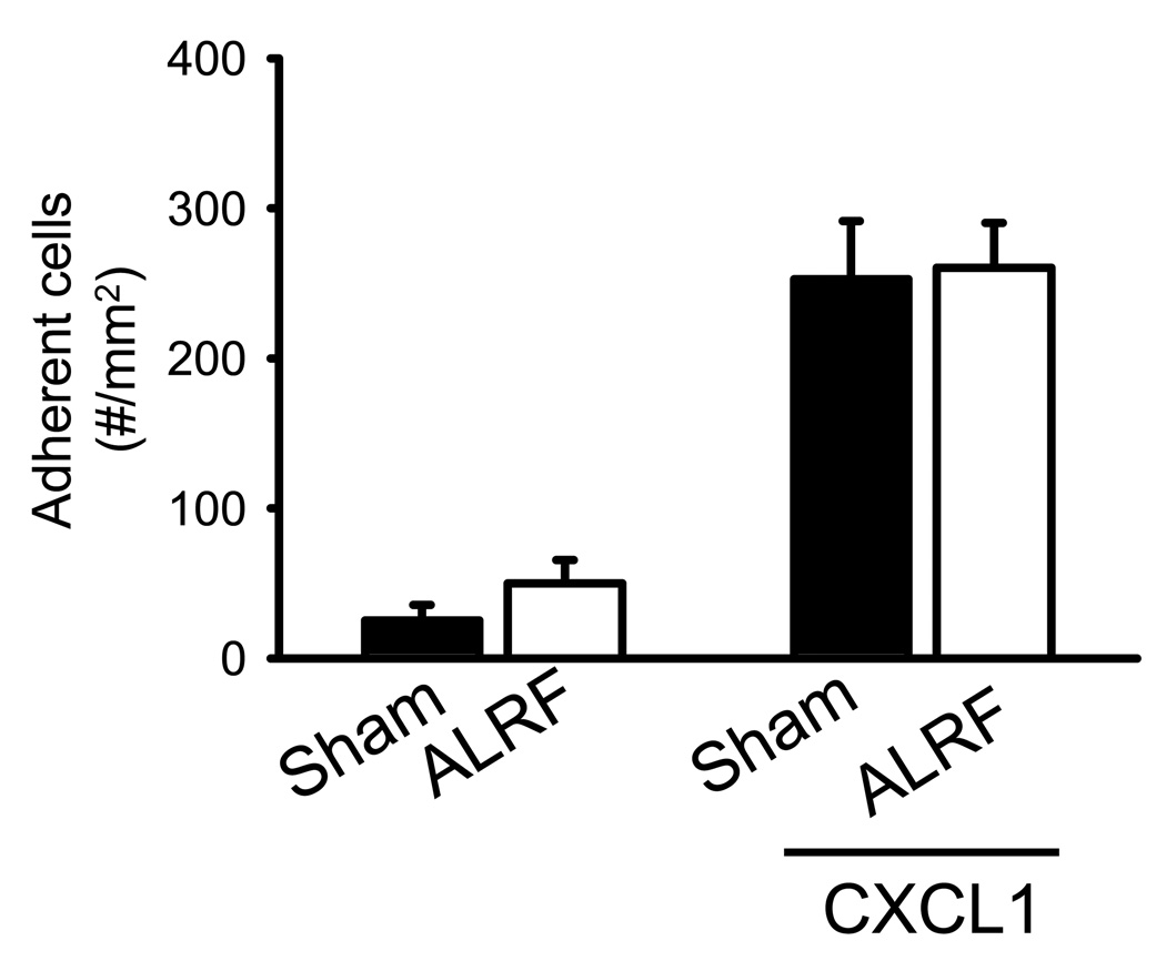
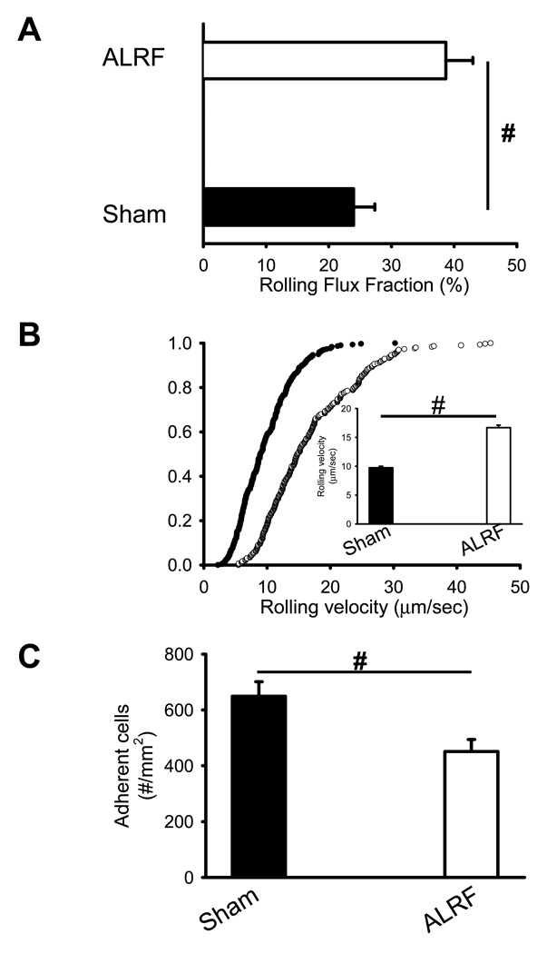
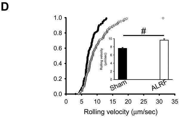
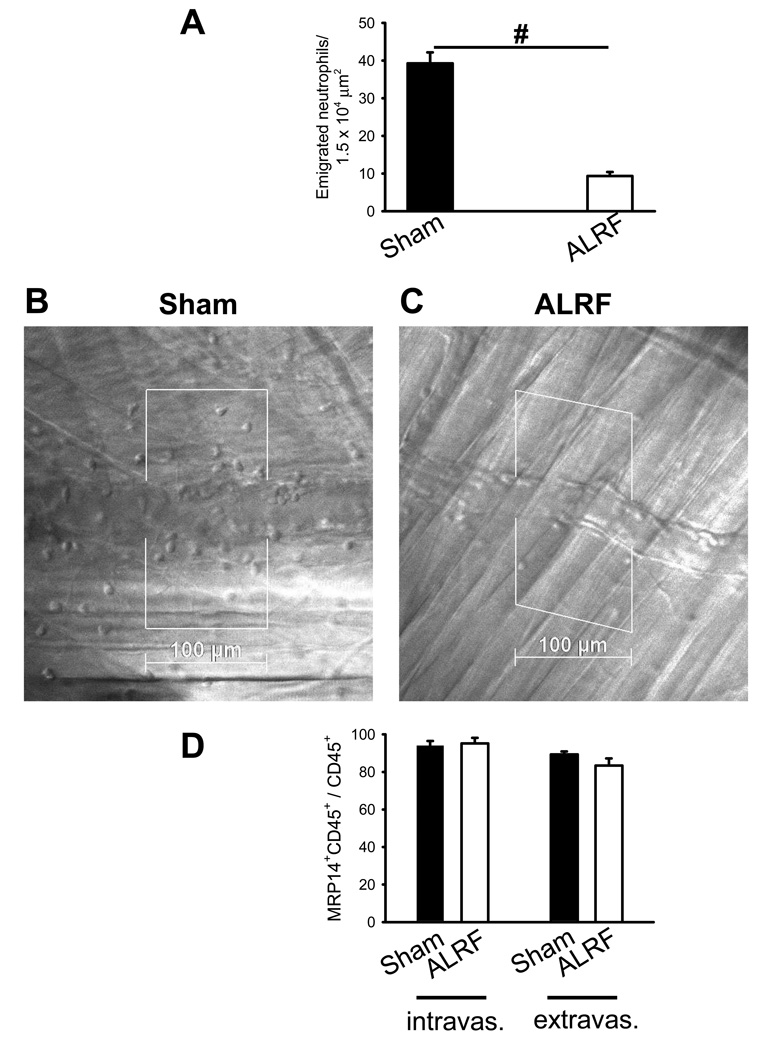
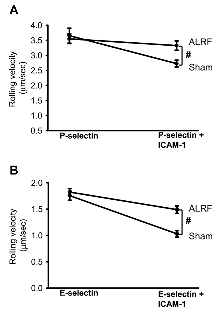
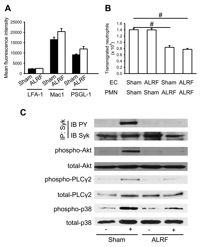
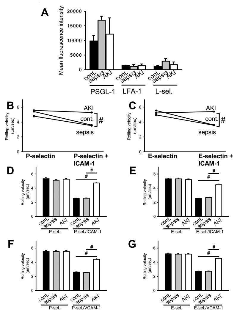
Acute loss of renal function reduces leukocyte recruitment into inflamed tissues, and we studied the molecular basis of this using intravital microscopy of cremaster muscle and an autoperfused flow chamber system after bilateral nephrectomy or sham operation in mice. Acute loss of renal function resulted in cessation of selectin-induced slow leukocyte rolling on E-selectin/intercellular adhesion molecule 1 (ICAM-1) and P-selectin/ICAM-1. It also reduced in vivo neutrophil extravasation (assessed by reflected light oblique transillumination) without affecting chemokine-induced arrest. This elimination of selectin-mediated slow leukocyte rolling was associated with a reduced phosphorylation of spleen tyrosine kinase, Akt, phospholipase C-γ2, and p38 MAPK. However, the levels of adhesion molecules located on the neutrophil surface were not altered. Leukocytes from critically ill patients with sepsis-induced acute kidney injury showed a significantly higher rolling velocity on E-selectin/ICAM-1- and P-selectin/ICAM-1-coated surfaces compared with patients with sepsis alone or healthy volunteers. Thus, an acute loss of renal function significantly impairs neutrophil rolling and transmigration, both in vivo and in vitro. These effects are due, in part, to decreased phosphorylation of selectin-dependent intracellular signaling pathways.


Conflict of interest statement

Disclosure

Conflict-of-interest disclosure: The authors declare no competing financial interests.

Figures

Figure 1
Figure 1. ALRF leads to inhibition of P-selectin mediated leukocyte activation in vivo
ALRF in mice was induced by bilateral nephrectomy 10 hours prior to intravital microscopy. (A) Leukocyte rolling flux fraction in untreated cremaster venules of mice with ALRF and sham-operated control mice (n=5). (B) Neutrophil rolling velocities in venules of mice with ALRF and sham-operated control mice. Data represent cumulative histograms of neutrophil rolling velocities measured after exteriorization of the cremaster muscle. The average rolling velocities (in µm/s) were calculated and are presented as means ± SEM (>100 neutrophils from 5 mice). (C) Numbers of adherent cells per mm2 in murine cremaster muscle venules. The cremaster muscle was exteriorized 10 h after bilateral nephrectomy or sham operation (n=5). # p < 0.05.
Figure 2
Figure 2. ALRF does not affect chemokine-induced arrest of leukocytes in vivo
Chemokine induced arrest of intravascular neutrophils in mice with ALRF and control mice was investigated after intravenous injection of 600ng CXCL1 by intravital microscopy of the cremaster muscle. The data was recorded and analyzed over the time period of 1 minute starting 15 seconds after the intravascular administration of CXCL1 (n=5). Data presented as means ± SEM.
Figure 3
Figure 3. ALRF alters the immune response during LPS-induced inflammation
6h after bilateral nephrectomy or sham operation, LPS was injected intrascrotally in order to induce inflammation. Intravital microscopy was performed 4h after LPS injection. (A) Leukocyte rolling flux fraction in inflamed cremaster venules 4h after LPS injection (n=5). (B) Neutrophil rolling velocities in inflamed venules of mice with ALRF and control mice. Data represent cumulative histograms of neutrophil rolling velocities measured 4h after LPS injection. The average rolling velocities (in µm/s) were calculated and are presented as means ± SEM (>100 neutrophils from 5 mice). (C) Numbers of adherent cells per mm2 in inflamed cremaster venules of mice with ALRF or control mice 4h after LPS injection (n=5). (D) After administration of a blocking antibody against P-selectin, the rolling velocity of neutrophils from mice with ALRF and sham operated control mice was determined. Data represent cumulative histograms of neutrophil rolling velocities measured 4h after LPS injection. The average rolling velocities (in µm/s) were calculated and are presented as means ± SEM (>100 neutrophils from 5 mice). # p < 0.05.
Figure 4
Figure 4. ALRF reduces neutrophil transmigration in vivo
Intravital microscopy was performed 10h after bilateral nephrectomy or sham operation. (A) Number of extravasated neutrophils in cremaster venules of LPS treated mice with ALRF or control mice per 1.5 × 104 µm2 tissue area. The measurements were performed 4 h after intrascrotal LPS injection (n=5). (B + C) Representative reflected light oblique transillumination microscopic pictures of cremaster muscle postcapillary venules of control mice (B) or mice with ALRF 4 h after LPS injection (C). Demarcations on each side of the venule determine the areas in which extravasated neutrophils were counted. # p < 0.05. (D) Mice received LPS intrascrotally 6 hours after bilateral nephrectomy or sham operation. 4 hours after LPS application, mice were sacrificed, the cremaster was removed, fixed, stained with PECAM1-, MRP14-, and CD45-antibodies and analyzed on a confocal microscope. The numbers of neutrophils (MRP14+CD45+) in the population of intravascular and extravascular leukocytes were counted and expressed as the percentage of MRP14+CD45+ cells within the CD45+ population (n=3).
Figure 5
Figure 5. ALRF completely abolishes selectin-mediated slow rolling
(A + B) The carotid artery of control mice and mice with ALRF were cannulated with a catheter, which was connected to autoperfused flow chambers. (A) Average rolling velocity of neutrophils on P-selectin (left) and P-selectin/ICAM-1 (right) is presented as mean ± SEM (n=6–10). (B) Average rolling velocity of neutrophils on E-selectin (left) and E-selectin/ICAM-1 (right) is presented as mean ± SEM (n=6–10). The wall shear stress in all flow chamber experiments was 5–6 dynes/cm2. # p < 0.05.
Figure 6
Figure 6. ALRF causes altered intracellular signaling and reduces transmigration in vitro
(A) Murine neutrophils were isolated from whole blood samples obtained from mice with ALRF and sham operated control mice and FACS analysis was performed to quantify the surface expression of PSGL-1, LFA-1 and Mac-1 (n=3). (B) Isolated neutrophils from mice with ALRF and sham operated control mice were allowed to transmigrate through bEnd.5 endothelial cells grown to confluence on a transwell filter in vitro. The endothelial cell layer was pretreated with plasma obtained from mice with ALRF or from sham operated control mice and the number of transmigrated neutrophils was determined (n=3). (C) Bone marrow–derived neutrophils were plated on uncoated (unstimulated) or E-selectin–coated wells for 10 minutes, and then lysates were prepared. Lysates were immunoprecipitated with anti-Syk, followed by immunoblotting (IB) with a general phosphotyrosine (PY, 4G10) antibody. Lysates were immunoblotted with antibody to phosphorylated PLCγ2 (phospho PLCγ2 (Tyr1217)), total PLCγ2 (n = 3), phosphorylated Akt (n = 3), total Akt (n = 3), phosphorylated p38 MAPK (phospho-p38), or total p38 (n = 3). # p < 0.05.
Figure 7
Figure 7. Selectin-mediated slow leukocyte rolling is abrogated in patients with sepsis-induced AKI
(A) Human neutrophils were isolated from whole blood samples obtained from patients presenting with sepsis or sepsis-induced AKI and healthy volunteers. FACS analysis was performed to quantify the surface expression of PSGL-1, LFA-1 and L-selectin on neutrophils (n=3–5). (B + C) The rolling velocity of neutrophils in whole blood samples from the three groups (healthy volunteers, sepsis, sepsis-induced AKI) was measured using microflow chambers coated with E-selectin or P-selectin alone and in combination with ICAM-1. (B) Average rolling velocity of neutrophils on P-selectin (left) and P-selectin/ICAM-1 (right) is presented as mean ± SEM (n=5). (C) Average rolling velocity of neutrophils on E-selectin (left) and E-selectin/ICAM-1 (right) is presented as mean ± SEM (n=5). (D + E) Human neutrophils were isolated from whole blood samples obtained from healthy volunteers and preincubated with control serum and serum obtained from patients with sepsis or sepsis-induced AKI for 30 minutes. The rolling velocity of neutrophils was measured using microflow chambers coated with E-selectin or P-selectin alone and in combination with ICAM-1. (D) Average rolling velocity of neutrophils on P-selectin and P-selectin/ICAM-1 is presented as mean ± SEM (n=3). (E) Average rolling velocity of neutrophils on E-selectin and E-selectin/ICAM-1 is presented as mean ± SEM (n=3). (F + G) Human monocytes were isolated from whole blood samples obtained from healthy volunteers and preincubated with control serum and serum obtained from patients with sepsis or sepsis-induced AKI for 30 minutes. The rolling velocity of monocytes was measured using microflow chambers coated with P-selectin (F) or E-selectin (G) alone and in combination with VCAM-1. (F) Average rolling velocity of monocytes on P-selectin and P-selectin/VCAM-1 is presented as mean ± SEM (n=3). (G) Average rolling velocity of monocytes on E-selectin and E-selectin/VCAM-1 is presented as mean ± SEM (n=3). The wall shear stress in all flow chamber experiments was 5–6 dynes/cm2. # p < 0.05.

Comment in

References

    1. Chertow GM, Burdick E, Honour M, et al. Acute kidney injury, mortality, length of stay, and costs in hospitalized patients. J Am Soc Nephrol. 2005;16:3365–3370. - PubMed
    1. Bagshaw SM, Uchino S, Bellomo R, et al. Septic acute kidney injury in critically ill patients: clinical characteristics and outcomes. Clin J Am Soc Nephrol. 2007;2:431–439. - PubMed
    1. Uchino S, Kellum JA, Bellomo R, et al. Acute renal failure in critically ill patients: a multinational, multicenter study. JAMA. 2005;294:813–818. - PubMed
    1. Thadhani R, Pascual M, Bonventre JV. Acute renal failure. N Engl J Med. 1996;334:1448–1460. - PubMed
    1. de Mendonca A, Vincent JL, Suter PM, et al. Acute renal failure in the ICU: risk factors and outcome evaluated by the SOFA score. Intensive Care Med. 2000;26:915–921. - PubMed

Publication types

LinkOut - more resources