. 2011 May 11;474(7350):235-8.
doi: 10.1038/nature09980.

Structure and function of a membrane component SecDF that enhances protein export

Affiliations

Structure and function of a membrane component SecDF that enhances protein export

Tomoya Tsukazaki et al. Nature. .

Abstract

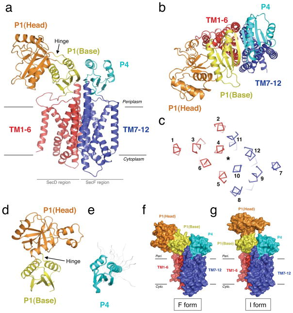
Protein translocation across the bacterial membrane, mediated by the secretory translocon SecYEG and the SecA ATPase, is enhanced by proton motive force and membrane-integrated SecDF, which associates with SecYEG. The role of SecDF has remained unclear, although it is proposed to function in later stages of translocation as well as in membrane protein biogenesis. Here, we determined the crystal structure of Thermus thermophilus SecDF at 3.3 Å resolution, revealing a pseudo-symmetrical, 12-helix transmembrane domain belonging to the RND superfamily and two major periplasmic domains, P1 and P4. Higher-resolution analysis of the periplasmic domains suggested that P1, which binds an unfolded protein, undergoes functionally important conformational changes. In vitro analyses identified an ATP-independent step of protein translocation that requires both SecDF and proton motive force. Electrophysiological analyses revealed that SecDF conducts protons in a manner dependent on pH and the presence of an unfolded protein, with conserved Asp and Arg residues at the transmembrane interface between SecD and SecF playing essential roles in the movements of protons and preproteins. Therefore, we propose that SecDF functions as a membrane-integrated chaperone, powered by proton motive force, to achieve ATP-independent protein translocation.


Figures

Figure 1
Figure 1. Structures of T. thermophilus SecDF
a, b, The crystal structure of full-length SecDF, viewed from the membrane side (a) and the periplasmic side (b). c, TSecDF cross-sectioned at the middle of the TM, viewed from the periplasm. The asterisk indicates the pseudo-symmetrical axis. TMs are numbered. d, Crystal structure of the P1 domain. e, NMR structure of the P4 domain after twenty superimpositions. The disordered regions are shown in gray. f, F form. Crystal structure of full-length SecDF. g, I form. The base subdomain of isolated P1 was docked onto that of the F form, as shown in f.
Figure 2
Figure 2. SecDF-dependent translocation completion
a, Identification of a SecDF- and PMF-dependent translocation step. SecDF-deficient (secD1) IMVs were incubated with 35S-labeled proOmpA(L59) to generate translocation intermediates. The protein translocation was continued in the presence or absence of ATP, PMF and DTT. b, Schematic depiction of the translocation intermediate of proOmpA(L59). c, Completion of the proOmpA(L59) translocation using IMVs from the secD1 (Cs) mutant expressing E. coli SecDF derivatives.
Figure 3
Figure 3. Functional charged residues and proton conduction of SecDF
a, Functionally important, conserved residues in the TM regions. The TMs are numbered. b, Complementation of the secD1 (Cs) growth and protein export defects by SecD/F mutants. c, Na+-dependent protein export by VSecDF-1. d, Na+-dependence of export (n=3). e, Single channel currents recorded by patch clamp in membrane patches excised from E. coli spheroplasts containing TSecDF or its mutants, and effects of a pH gradient and casein. f, The channel open probability (n=6 with casein, n=4 without casein).
Figure 4
Figure 4. A working model of the PMF-driven translocation enhancement by SecDF
a, F form, capturing state. b, I form, holding state. c, I to F transition and substrate-releasing state. The two essential charged residues of ESecDF are highlighted. SecDF is colored as in Fig. 2. SecYEG, gray; SecA, green; pre-protein, black line; proton movement, white arrow. See the main text discussion for details.

References

    1. van den Berg B, et al. X-ray structure of a protein-conducting channel. Nature. 2004;427:36–44. - PubMed
    1. Zimmer J, Nam Y, Rapoport TA. Structure of a complex of the ATPase SecA and the protein-translocation channel. Nature. 2008;455:936–43. - PMC - PubMed
    1. Tsukazaki T, et al. Conformational transition of Sec machinery inferred from bacterial SecYE structures. Nature. 2008;455:988–91. - PMC - PubMed
    1. du Plessis DJ, Nouwen N, Driessen AJ. The Sec translocase. Biochim Biophys Acta. 2011;1808:851–65. - PubMed
    1. Driessen AJ, Wickner W. Proton transfer is rate-limiting for translocation of precursor proteins by the Escherichia coli translocase. Proc Natl Acad Sci U S A. 1991;88:2471–5. - PMC - PubMed

Publication types

MeSH terms

LinkOut - more resources