. 2011 Jul;20(7):1231-43.
doi: 10.1002/pro.651. Epub 2011 Jun 14.

Site-directed spin labeling electron paramagnetic resonance study of the ORF1 protein from a mouse L1 retrotransposon

Affiliations

Site-directed spin labeling electron paramagnetic resonance study of the ORF1 protein from a mouse L1 retrotransposon

Kurt Januszyk et al. Protein Sci. 2011 Jul.

Abstract

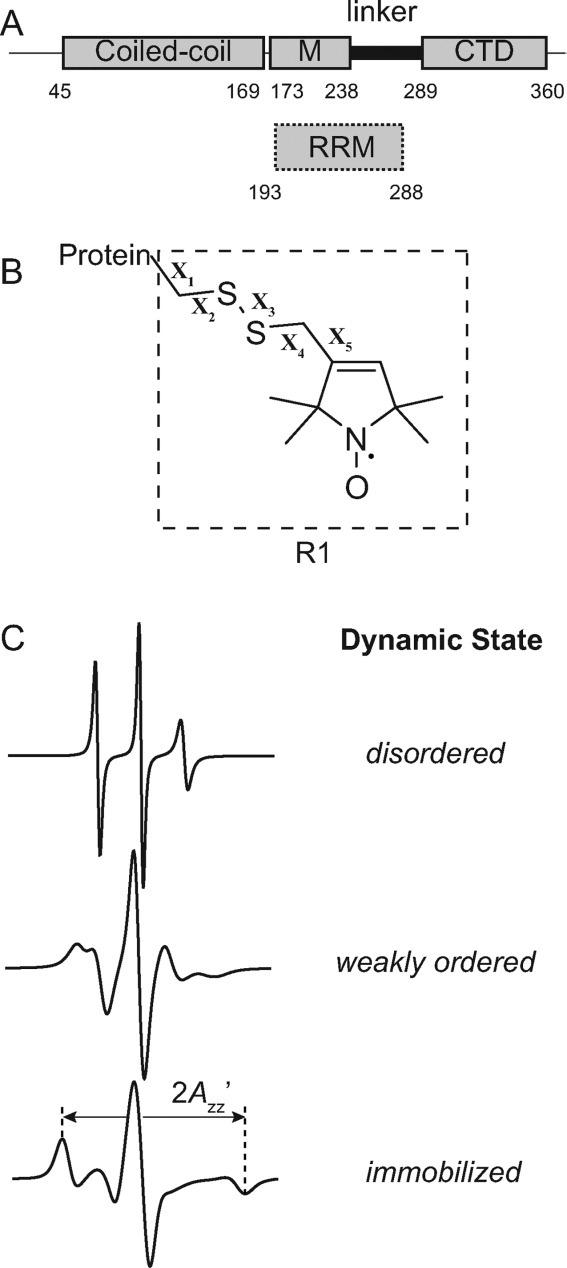
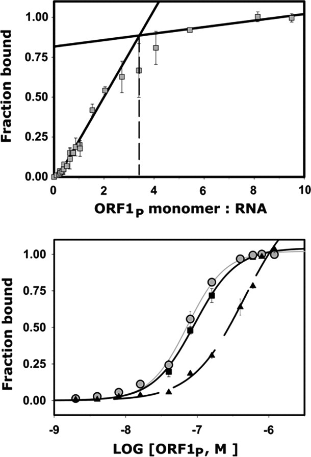
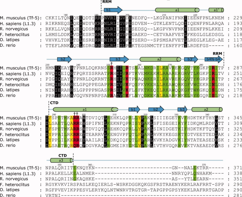
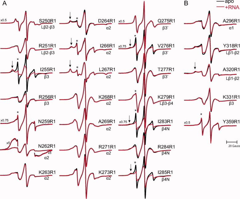
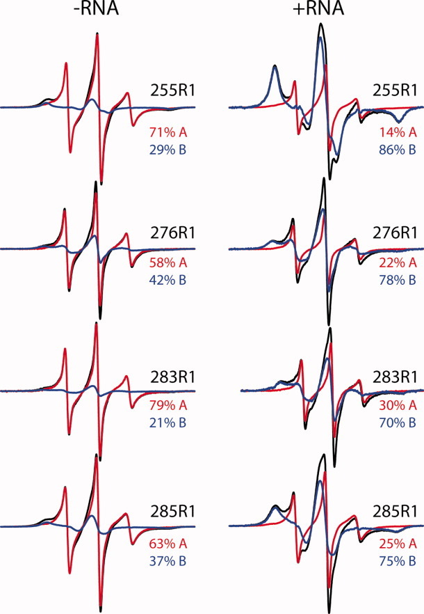
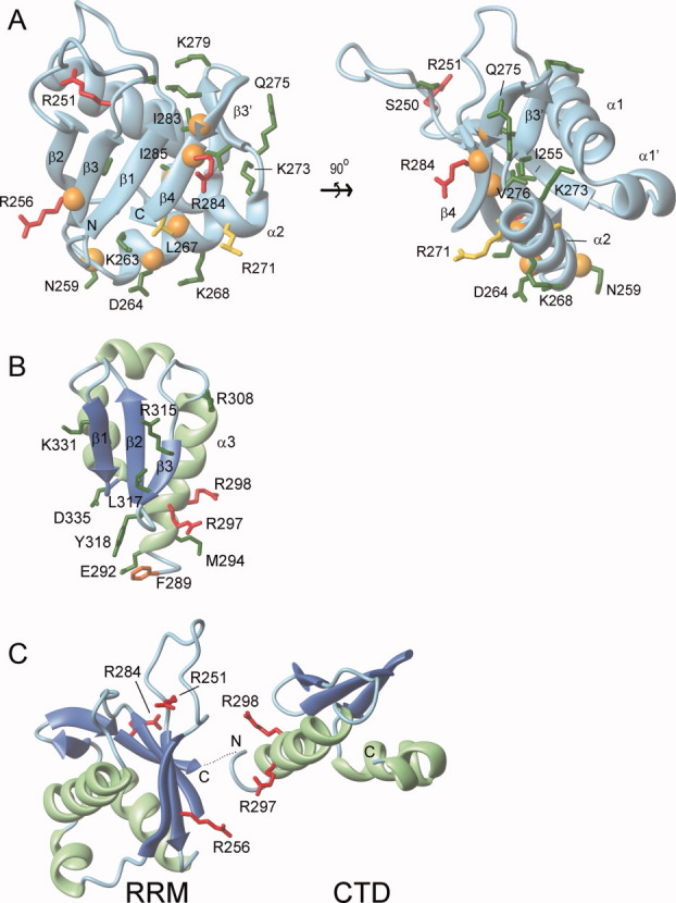
Long interspersed nuclear element-1 is a highly abundant mammalian retrotransposon that comprises 17% of the human genome. L1 retrotransposition requires the protein encoded by open reading frame-1 (ORF1p), which binds single-stranded RNA with high affinity and functions as a nucleic acid chaperone. ORF1p has been shown to adopt a homo-trimeric, asymmetric dumbbell-shaped structure. However, its atomic-level structure and mechanism of RNA binding remains poorly understood. Here, we report the results of a site-directed spin labeling electron paramagnetic resonance (SDSL-EPR) study of 27 residues within the RNA binding region of the full-length protein. The EPR data are compatible with the large RNA binding lobe of ORF1p containing a RNA recognition motif (RRM) domain and a carboxyl-terminal domain (CTD) that are predicted from crystallographic and NMR studies of smaller fragments of the protein. Interestingly, the EPR data indicate that residues in strands β3 and β4 of the RRM are structurally unstable, compatible with the previously observed sensitivity of this region to proteolysis. Affinity measurements and RNA-dependent EPR spectral changes map the RNA binding site on ORF1p to residues located in strands β3 and β4 of the RRM domain and to helix α1 of the CTD. Complementary in vivo studies also identify residues within the RRM domain that are required for retrotransposition. We propose that in the context of the full-length trimeric protein these distinct surfaces are positioned adjacent to one another providing a continuous surface that may interact with nucleic acids.


Figures

Figure 1
Figure 1
Schematic of ORF1p, the R1 spin label, and representative EPR spectra. (A) The top image displays a schematic of the ORF1p protein from the mouse L1 retrotransposon. Regions of the protein previously shown to be partially resistant to trypsin digestion are indicated by rectangles and correspond to a coiled-coil region, a middle region (M) and a C-terminal domain (CTD). The segment connecting the CTD and middle region is susceptible to trypsin proteolysis and is referred to as the linker. The lower image indicates where the presumed RNA recognition motif (RRM) binding domain is located based on the recently determined crystal structure of this domain from the human ORF1p protein (hORF1p). The amino acid number defining the beginning and end of each element is indicated. (B) Structure of the R1 side chain showing the dihedral angle designations (X1–X5). (C) Simulated EPR spectra corresponding to three fundamental dynamic modes of the R1 side chain in proteins: disordered (top trace), weakly ordered (middle trace), and immobilized (lower trace). Simulations were carried out using the NLSL.MOMD program available at (http://www.acert.cornell.edu/index_files/acert_ftp_links.php). In each case, the order parameter (S) and correlation time (τ) for the motion are given in the format {S,τ}. Details of spectral simulations are presented by Columbus et al.
Figure 2
Figure 2
Representative RNA binding data from the full length and mutant ORF1p proteins. (A) Binding assay used to determine the stoichiometry of the complex between full-length ORF1p and a 60-mer L1 RNA molecule. The results of a double filter nitrocellulose binding assay using near equal molar concentrations of each component is shown. The black lines plot the linear regression for data points in the linear and saturation phases of the binding curve. Extrapolation (dotted lines) revealed ∼1.1 ORF1p trimers bind to a single RNA molecule. (B) Representative RNA binding assays of wild-type and mutant ORF1p proteins. The strength of RNA binding to a 60-mer L1 RNA was tested. Shown is data for: wild-type ORF1p (black squares, black line), and Arg256Cys/Cys174Arg (gray circles, gray lines), and Asn262Cys/Cys174Arg (black triangles, black dashed line) mutants. The data were fit to a binding model that assumed 1:1 stoichiometry and yielded KD values of 92 ± 9 nM, 47 ± 16 nM, and >1500 nM, respectively. A complete list of the binding data is presented in Table I.
Figure 3
Figure 3
Primary sequence alignment of ORF1 proteins and a summary of the RNA binding and in vivo results. The alignment of amino acids of ORF1p contains the RRM identified by crystallography and the CTD identified by NMR. Amino acids that were mutated to cysteine or alanine residues and determined for their ability to bind a 60-mer L1 RNA molecule are color coded: green (KD < 3X wild type), yellow (3X wild type ≤ KD ≤ 6.1X wild type), and red (KD > 6.1X wild type). Residues that are highly conserved but were not mutated are colored gray. Mutants that disrupted in vivo retrotransposition are indicated by daggers. The ORF1 protein alignments were generated for mouse (from the Tf-5 retrotransposon, AAC53541.1), human (from the human L1.3 retrotransposon, AAB59367), rat (S21345), mumichog (AF055640), Zebrafish (CAD61093), and medaka (AAS83199) using CLUSTALW.
Figure 4
Figure 4
EPR spectra of apo and RNA bound mouse ORF1p. EPR spectra of ORF1p R1 mutants at sites in the RRM (panel A) or CTD (panel B) in the absence (black) and a presence of a 60-mer L1 RNA (red). Spectral features indicating highly dynamic components are designated by an asterisk; those indicating relatively immobilized states are designated by an arrow. The magnetic field scan width is 100 G. All spectra are normalized to the same concentration of nitroxide. Vertical scaling factors (left side of spectrum) were applied to select spectra for display purposes.
Figure 5
Figure 5
Individual dynamic components of Ile255R1, Val276R1, Ile283R1, and Ile285R1 spectra. EPR spectra of the indicated ORF1p mutants with (right panel) or without RNA (left panel) shown as black traces. For each mutant in the presence or absence of RNA, the EPR spectrum is modeled as a weighted sum of the same two dynamic components (red, blue traces), with the percentage of each component present given on the right. In the presence of RNA, an increase in the relatively immobilized component and corresponding decrease in the highly dynamic component is evident.
Figure 6
Figure 6
RNA binding model for the RRM and CTD. Residues important for RNA binding within the (A) RRM domain and (B) CTD. Amino acids within ORF1p that were mutated to cysteine or alanine residues and determined for their ability to bind a 60-mer L1 RNA molecule are color coded: green (KD < 3X wild type), yellow (3X wild type ≤ KD ≤ 6X wild type), and red (KD > 6X wild type). An orange sphere positioned at the alpha carbon atom indicates R1 labeled cysteine mutants that exhibited RNA-dependent changes in their EPR spectra. (C) Proposed primary interaction surface with RNA. The CTD and RRM domain are positioned adjacent to one to form a continuous surface that interacts with single stranded RNA. It is unclear whether this surface is formed by the RRM and CTD from a single monomer or from different monomers within the trimer. The orientation of the RNA molecule on this surface is also unknown. In panels B and C, ribbon diagrams of the RRM domain and CTD are shown with the sheet and helices colored blue and green, respectively. The secondary structure topology is labeled accordingly for the RRM (PDB 2w7a) and the CTD (PDB 2jrb).

References

    1. Goodier JL, Kazazian HH., Jr Retrotransposons revisited: the restraint and rehabilitation of parasites. Cell. 2008;135:23–35. - PubMed
    1. Babushok DV, Kazazian HH., Jr. Progress in understanding the biology of the human mutagen LINE-1. Hum Mutat. 2007;28:527–539. - PubMed
    1. Han JS, Boeke JD. LINE-1 retrotransposons: modulators of quantity and quality of mammalian gene expression? Bioessays. 2005;27:775–784. - PubMed
    1. Kazazian HH, Jr, Moran JV. The impact of L1 retrotransposons on the human genome. Nat Genet. 1998;19:19–24. - PubMed
    1. Ostertag EM, Kazazian HH. Biology of mammalian L1 retrotransposons. Annu Rev Genet. 2001;35:501–538. - PubMed

Publication types

LinkOut - more resources