. 2011 Jul;20(7):1244-55.
doi: 10.1002/pro.652.

Probing the interactions of an acyl carrier protein domain from the 6-deoxyerythronolide B synthase

Affiliations

Probing the interactions of an acyl carrier protein domain from the 6-deoxyerythronolide B synthase

Louise K Charkoudian et al. Protein Sci. 2011 Jul.

Abstract

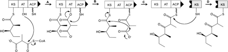
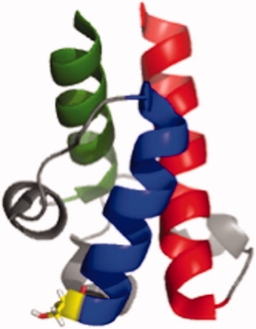
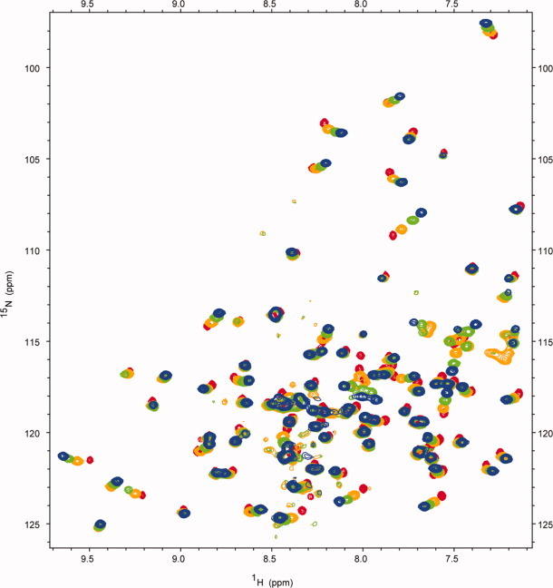
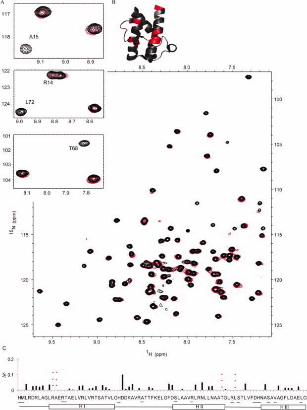
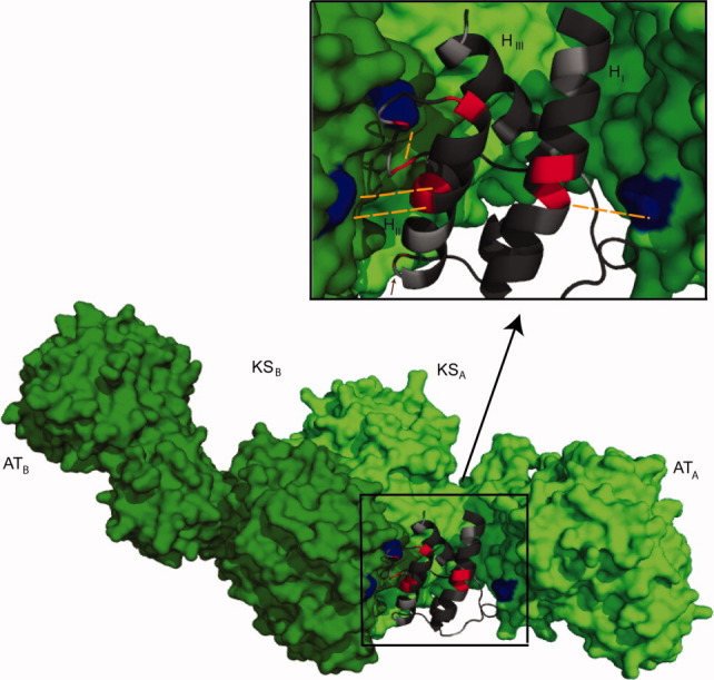
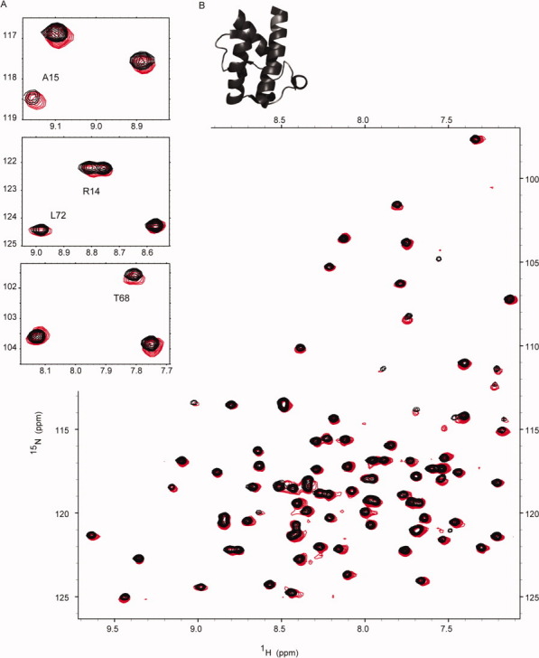
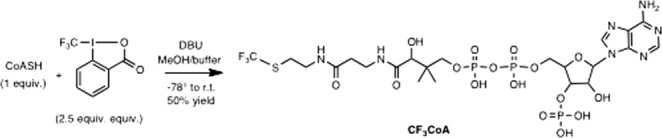
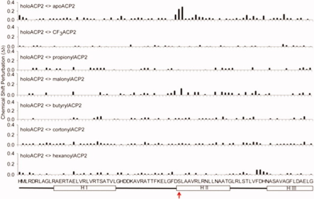
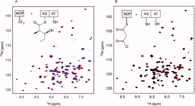
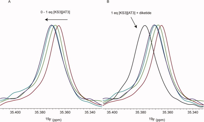
The assembly-line architecture of polyketide synthases (PKSs) provides an opportunity to rationally reprogram polyketide biosynthetic pathways to produce novel antibiotics. A fundamental challenge toward this goal is to identify the factors that control the unidirectional channeling of reactive biosynthetic intermediates through these enzymatic assembly lines. Within the catalytic cycle of every PKS module, the acyl carrier protein (ACP) first collaborates with the ketosynthase (KS) domain of the paired subunit in its own homodimeric module so as to elongate the growing polyketide chain and then with the KS domain of the next module to translocate the newly elongated polyketide chain. Using NMR spectroscopy, we investigated the features of a structurally characterized ACP domain of the 6-deoxyerythronolide B synthase that contribute to its association with its KS translocation partner. Not only were we able to visualize selective protein-protein interactions between the two partners, but also we detected a significant influence of the acyl chain substrate on this interaction. A novel reagent, CF₃-S-ACP, was developed as a ¹⁹F NMR spectroscopic probe of protein-protein interactions. The implications of our findings for understanding intermodular chain translocation are discussed.


Figures

Figure 1
Figure 1
Catalytic cycle of a minimal module in a multimodular PKS assembly line. The core catalytic module comprises three types of domains: AT, acyl transferase; ACP, acyl carrier protein; and KS, ketosynthase. The Ppant prosthetic group of the ACP domain is designated with a curly line. Noncovalent protein–protein interfaces lying outside the core catalytic module influence the selectivity of intermodular chain translocation and are shown as shape-complementary tabs. The AT recognizes an α-carboxylated Coenzyme A thioester (e.g., methylmalonyl-CoA) and transfers this extender unit onto the Ppant arm of the ACP domain via a transacylation reaction involving a Ser residue in its active site (A and B). The acylated KS and ACP domains then collaborate to elongate the polyketide chain by two carbons via a decarboxylative condensation reaction (C). The elongated polyketide is transferred from the ACP of the donor module to the KS of the recipient module (gray) via thioester exchange (D). Auxiliary enzyme domains are frequently found in PKS modules and are responsible for further modification of the resulting β-ketothioester; these domains are not shown for simplicity.
Figure 2
Figure 2
Solution structure of ACP2. The tertiary fold of ACP2 is a three-helix bundle (helix I = red, helix II = blue, and helix III = green). The Ppant arm is covalently attached to S54 (yellow) at the N-terminus of helix II.
Figure 3
Figure 3
Superimposed [1H, 15N]-HSQC spectra of uniformly 15N-labeled apo-ACP2 at pH 5.5 (red), 6.0 (orange), 6.5 (green), and 7.0 (blue).
Figure 4
Figure 4
Spectroscopic changes observed upon titration of uniformly 15N-labeled ACP2 with chain translocation partner [KS3][AT3]. (A) Superimposed [1H, 15N]-HSQC spectra of uniformly 15N-labeled apo-ACP2 (black) and apo-ACP2 mixed with 0.75 equiv of [KS3][AT3] (red). Insets show the disappearance of the R14, A15, T68, and L72 [1H, 15N]-HSQC peaks of apo-ACP2 upon titration of [KS3][AT3]. (B) Residues whose HSQC peaks decrease in intensity by >60% upon titration of [KS3][AT3] are highlighted on the ACP2 structure. Those that could not be assigned at pH 7 are tinted light gray. (C) Chemical shift perturbations (Δδ) of apo-ACP2 alone versus apo-ACP2 in the presence of the [KS3][AT3] protein. ACP2 residues that could not be assigned an [1H, 15N]-HSQC peak at pH 7.0 are underlined; residues that disappear in the presence of the [KS3][AT3] protein are denoted with red “***.”
Figure 5
Figure 5
Proposed docking model for the structurally characterized ACP2 (dark gray) onto [KS3][AT3] (green) during the chain transfer mode of interaction. Apo-ACP2 residues with solvent-exposed side chains whose backbone HSQC peaks decrease in intensity by >60% upon addition of [KS3][AT3] are highlighted in red (L24, R26, N62, R61, T68, and T75). Salt bridges and hydrogen-bonding interactions between the ACP2 residue (red) and [KS3][AT3] partner residue (blue) are highlighted in orange. ACP2 residues that could not be assigned at pH 7 are tinted light gray.
Figure 6
Figure 6
Superimposed [1H, 15N]-HSQC spectra of uniformly 15N-labeled apo-ACP2 (black) and ACP2 mixed with 0.75 equiv of [KS5][AT5] (red). Insets highlight the same regions as in Figure 4 but show that the [1H, 15N]-HSQC peaks of R14, A15, T68, and L72 from ACP2 do not disappear upon titration of the [KS5][AT5]. Under these conditions, none of the ACP2 residues disappeared or moved significantly. (B) Accordingly, the ACP2 structure only represents in light gray the residues that could not be assigned at pH 7.
Scheme 1
Scheme 1
Preparation of the S-trifluoromethylated Coenzyme A.
Figure 7
Figure 7
Comparison of the [1H, 15N]-HSQC spectra of selected ACP2 analogs. Chemical shift perturbations (Δδ) of holo-ACP2 versus apo-, trifluoromethyl-S,- and five acyl- (propionyl, malonyl, butyryl, crotonyl, and hexanoyl) ACP2s. The primary sequence of ACP2 and the positions of the α-helices are indicated. The attachment site of the Ppant arm (S54) is noted with a red arrow.
Figure 8
Figure 8
(A) Superimposed [1H, 15N]-HSQC spectra of uniformly 15N-labeled CF3-S-ACP2 titrated with 0.75 equiv of [KS3][AT3] at pH 7.0 (red) versus uniformly 15N-labeled CF3-S-ACP2 titrated with 0.75 equiv of [KS3][AT3] and excess (2S,3R)-2-methyl-3-hydroxypentanoyl-N-acetylcysteamine thioester at pH 7.0 (blue). (B) Superimposed [1H, 15N]-HSQC spectra of uniformly 15N-labeled holo-ACP2 titrated with 0.75 equiv of [KS3][AT3] (red) versus uniformly 15N-labeled malonyl-ACP2 titrated with 0.75 equiv of [KS3][AT3] at pH 7.0 (black). A schematic representation of the protein complexes evaluated is outlined in the top left box of each spectrum.
Figure 9
Figure 9
19F NMR of CF3-S-ACP2 in the absence and presence of [KS3][AT3] and diketide-acylated [KS3][AT3]. (A) 19F NMR of CF3-S-ACP2 alone and in the presence of 0.25 equiv [KS3][AT3], 0.5 equiv [KS3][AT3], 0.75 equiv [KS3][AT3], and 1 equiv [KS3][AT3]. (B) 19F NMR of CF3-S-ACP2 in the presence of diketide-acylated [KS3][AT3].

Similar articles

Cited by

References

    1. Khosla C, Tang Y, Chen AY, Schnarr NA, Cane DE. Structure and mechanism of the 6-deoxyerythronolide B synthase. Annu Rev Biochem. 2007;76:195–221. - PubMed
    1. Weissman KJ, Leadlay PF. Combinatorial biosynthesis of reduced polyketides. Nat Rev Microbiol. 2005;3:925–936. - PubMed
    1. Fischbach MA, Walsh CT. Assembly-line enzymology for polyketide and nonribosomal peptide antibiotics: logic, machinery, and mechanisms. Chem Rev. 2006;106:3468–3496. - PubMed
    1. McDaniel R, Welch M, Hutchinson CR. Genetic approaches to polyketide antibiotics. Chem Rev. 2005;105:543–558. - PubMed
    1. Khosla C, Kapur S, Cane DE. Revisiting the modularity of modular polyketide synthases. Curr Opin Chem Biol. 2009;13:135–143. - PMC - PubMed

Publication types

MeSH terms

LinkOut - more resources