

Segmentations of MRI images of the female pelvic floor: a study of inter- and intra-reader reliability
- PMID: 21563253
- PMCID: PMC4364418
- DOI: 10.1002/jmri.22478
Segmentations of MRI images of the female pelvic floor: a study of inter- and intra-reader reliability
Abstract
Purpose: To describe the inter- and intra-operator reliability of segmentations of female pelvic floor structures.
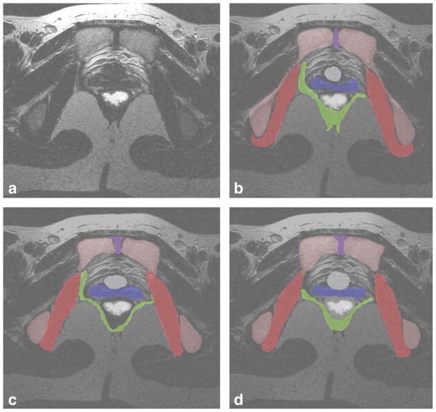
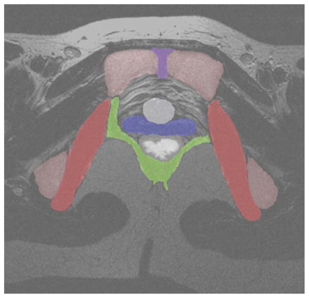
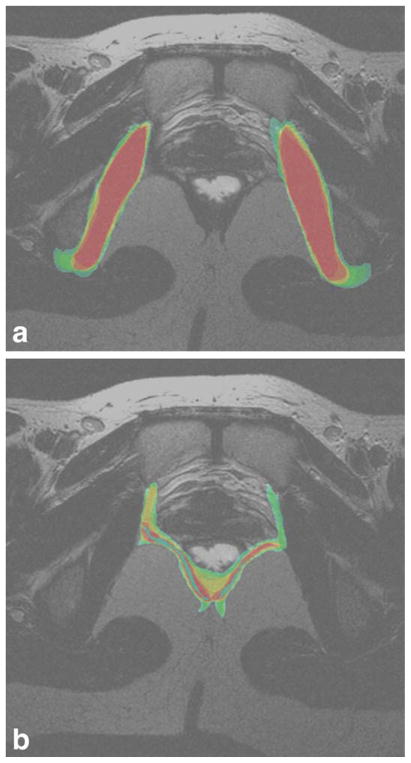
Materials and methods: Three segmentation specialists were asked to segment out the female pelvic structures in 20 MR datasets on three separate occasions. The STAPLE algorithm was used to compute inter- and intra-segmenter agreement of each organ in each dataset. STAPLE computed the sensitivity, specificity, and positive predictive values (PPV) for inter- and intra-segmenter repeatability. These parameters were analyzed using intra-class correlation analysis. Correlation of organ volume to PPV and sensitivity was also computed.
Results: Mean PPV of the segmented organs ranged from 0.82 to 0.99, and sensitivity ranged from 33 to 96%. Intra-class correlation ranged from 0.07 to 0.98 across segmenters. Pearson correlation of volume to sensitivity were significant across organs, ranging from 0.54 to 0.91. Organs with significant correlation of PPV to volume were bladder (-0.69), levator ani (-0.68), and coccyx (-0.63).
Conclusion: Undirected manual segmentation of the pelvic floor organs are adequate for locating the organs, but poor at defining structural boundaries.
Copyright © 2011 Wiley-Liss, Inc.
Figures
References
-
- Hoyte L, Thomas J, Foster RT, Shott S, Jakab M, Weidner AC. Racial differences in pelvic geometry among asymptomatic nulliparas as seen on three-dimensional MR images. Proceedings of the Annual Meeting of Society of Gynecologic Surgeons; Rancho Mirage, California. 2005.
-
- Cornella JL, Hibner M, Fenner DE, Kriegshauser JS, Hentz J, Magrina JF. Three-dimensional reconstruction of magnetic resonance images of the anal sphincter and correlation between sphincter volume and pressure. Am J Obstet Gynecol. 2003;189:130–135. - PubMed
-
- Hoyte L, Schierlitz L, Zou K, Flesh G, Fielding JR. Two and 3 dimensional MRI comparison of Levator Ani structure, volume and integrity in women with stress incontinence and prolapse. Am J Obstet Gynecol. 2001;185:11–19. - PubMed
-
- Cline HE, Lorensen WE, Ludke S, Crawford CR, Teeter BC. Two algorithms for the three-dimensional reconstruction of tomograms. Med Phys. 1988;15:320–327. - PubMed
-
- Kikinis R, Gleason PL, Moriarty TM, et al. Computer-assisted interactive three-dimensional planning for neurosurgical procedures. Neurosurgery. 1996;38:640–649. discussion 649–651. - PubMed
Publication types
MeSH terms
Grants and funding
- U10 HD041261/HD/NICHD NIH HHS/United States
- U01 HD041249/HD/NICHD NIH HHS/United States
- U10 HD41261/HD/NICHD NIH HHS/United States
- U10 HD41248/HD/NICHD NIH HHS/United States
- U10 HD41250/HD/NICHD NIH HHS/United States
- U10 HD41268/HD/NICHD NIH HHS/United States
- U10 HD041263/HD/NICHD NIH HHS/United States
- U10 HD041267/HD/NICHD NIH HHS/United States
- U10 HD041248/HD/NICHD NIH HHS/United States
- UG1 HD041267/HD/NICHD NIH HHS/United States
- U10 HD041269/HD/NICHD NIH HHS/United States
- U10 HD041268/HD/NICHD NIH HHS/United States
- U10 HD041250/HD/NICHD NIH HHS/United States
- U10 HD41267/HD/NICHD NIH HHS/United States
- U10 HD41269/HD/NICHD NIH HHS/United States
- U10 HD41263/HD/NICHD NIH HHS/United States
- U01 HD41249/HD/NICHD NIH HHS/United States
