. 2012 Mar;16(3):605-15.
doi: 10.1111/j.1582-4934.2011.01338.x.

E2F-1- and E2Ftr-mediated apoptosis: the role of DREAM and HRK

Affiliations

E2F-1- and E2Ftr-mediated apoptosis: the role of DREAM and HRK

Hongying Hao et al. J Cell Mol Med. 2012 Mar.

Abstract

E2F-1-deleted mutant, 'truncated E2F' (E2Ftr, E2F-1[1-375]), lacking the carboxy-terminal transactivation domain, was shown to be more potent at inducing cancer cell apoptosis than wild-type E2F-1 (wtE2F-1; full-length E2F-1). Mechanisms by which wtE2F-1 and E2Ftr induce apoptosis, however, are not fully elucidated. Our study demonstrates molecular effects of pro-apoptotic BH3-only Bcl-2 family member Harakiri (Hrk) in wtE2F-1- and E2Ftr-induced melanoma cell apoptosis. We found that Hrk mRNA and Harakiri (HRK) protein expression was highly up-regulated in melanoma cells in response to wtE2F-1 and E2Ftr overexpression. HRK up-regulation did not require the E2F-1 transactivation domain. In addition, Hrk gene up-regulation and HRK protein expression did not require p53 in cancer cells. Hrk knockdown by Hrk siRNA was associated with significantly reduced wtE2F-1- and E2Ftr-induced apoptosis. We also found that an upstream factor, 'downstream regulatory element antagonist modulator' (DREAM), may be involved in HRK-mediated apoptosis in response to wtE2F-1 and E2Ftr overexpression. DREAM expression levels increased following wtE2F-1 and E2Ftr overexpression. Western blotting detected increased DREAM primarily in dimeric form. The homodimerization of DREAM resulting from wtE2F-1 and E2Ftr overexpression may contribute to the decreased binding activity of DREAM to the 3'-untranslated region of the Hrk gene as shown by electromobility shift assay. Results showed wtE2F-1- and E2Ftr-induced apoptosis is partially mediated by HRK. HRK function is regulated in response to DREAM. Our findings contribute to understanding the mechanisms that regulate wtE2F-1- and E2Ftr-induced apoptosis and provide insights into the further evaluation of how E2Ftr-induced apoptosis may be used for therapeutic gain.


Figures

Fig 1
Fig 1
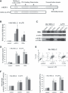
Overexpression of wtE2F-1 and E2Ftr leads to increased Hrk expression and apoptosis in melanoma cells. (A) Schematic representation of wtE2F-1 and E2Ftr domain structure. Two melanoma cell lines (SK-MEL-2 and A375) were infected with Ad-LacZ (control vector), Ad-wtE2F-1 or Ad-E2Ftr at MOI 100. After 24 hrs of infection, real-time RT-PCR (B) and Western blot analysis (C) were performed as described in Materials and methods. (B) RT-PCR results are expressed as Hrk fold increase relative to control vector-infected cells adjusted for α-actin as an internal control. Each experiment is a representation of three independent experiments performed in duplicate (bars: mean ± S.D.; *P < 0.05 compared with control; n = 3). (C) Cell lysates were subjected to Western blot analysis using HRK, wtE2F-1 or E2Ftr and α-actin antibody. (D) After 48 hrs of infection, the percentage of apoptotic cells showing typical apoptotic nuclei by Hoechst 33258 staining was counted under a fluorescence microscope. The percentage of apoptotic cells (annexin V-PE+ cells) were also determined by flow cytometry analysis (E) after 48 hrs of infection, as described in Materials and methods. (F) After 48 hrs of designated infection, the OD405 values of the cell lysates were measured as caspase-9 activity. The results were expressed as the fold change in treated cells over that of the control cells. Values represent mean ± S.D. of three independent experiments (bars: mean ± S.D.; **P < 0.01 compared with control; n = 3).
Fig 2
Fig 2
Up-regulation of HRK in wtE2F-1- and E2Ftr-induced apoptosis is independent of p53 status. H1299 and SAOS2 cells, which lack p53, were infected at MOI 100. After 24 hrs of infection, RT-PCR (A) and Western blot analysis (B) were performed as described in Figure 1B and C. After 48 hrs of infection, the percentage of apoptotic cells was counted by Hoechst 33258 staining under a fluorescence microscope (C) and determined by annexin V-PE staining by flow cytometry analysis (D). Values represent mean ± S.D. of three independent experiments (bars: mean ± S.D.; **P < 0.01 compared with control; n = 3).
Fig 3
Fig 3
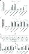
Hrk knockdown by siRNA was associated with significantly reduced wtE2F-1- or E2Ftr-induced apoptosis. SK-MEL-2 and A375 cells were infected with control vector, Ad-wtE2F-1 or Ad-E2Ftr at MOI 100, followed by transfection with control siRNA or Hrk siRNA as indicated. (A) RT-PCR was performed after 24 hrs of transfection as described in Materials and methods. The bar graph is expressed as Hrk fold increase relative to control vector-infected cells adjusted for α-actin as an internal control. Each experiment is a representation of three independent experiments performed in duplicate (Ctrl: control; bars: mean ± S.D.; *P < 0.05; **P < 0.01 compared with either wtE2F-1 or E2Ftr infection plus control siRNA transfection; n = 3). After 48 hrs of transfection, the percentage of cells showing typical apoptotic nuclei was counted by Hoechst 33258 staining under a fluorescence microscope (B) and determined by annexin V-PE staining by flow cytometry analysis (C). (D) After 48 hrs of transfection, caspase-9 activity was determined as in Figure 1F. Values represent mean ± S.D. of three independent experiments. (Ctrl: control; bars: mean ± S.D.; *P < 0.05; **P < 0.01 compared with either wtE2F-1 or E2Ftr infection plus control siRNA transfection; n = 3).
Fig 4
Fig 4
wtE2F-1 and E2Ftr are not directly acting to induce apoptosis at mitochondria. After 24 hrs of infection, A375 cells (A) and SK-MEL-2 cells (B) were stained with mitotracker red 580 and HRK (A) or E2F-1 (B) and then counterstained with DAPI (blue) (upper left panel) as described in Materials and methods (magnification, ×600). (A) An overlay (lower right panel) of the red (mitochondria, lower left panel) and the green (HRK, upper right panel) is provided in yellow to show co-localization of HRK in mitochondria. (B) wtE2F-1 or E2Ftr is primarily distributed in the nuclei and cytoplasma as shown in green (right upper panel). An overlay (lower right panel) of the red (mitochondria, lower left panel) and the green (wtE2F-1 or E2Ftr, right upper panel) is not shown in yellow, which suggests wtE2F-1 or E2Ftr is not located in mitochondria. Insets represent higher magnifications of the boxed areas (bar = 20μm).
Fig 5
Fig 5
wtE2F-1 and E2Ftr expression resulted in reduced binding of DREAM to the Hrk gene and was correlated with increased HRK up-regulation and apoptosis. SK-MEL-2 and A375 cells were infected as indicated. After 24 hrs of infection, nuclear and cytoplasma protein were extracted. (A) Nuclear proteins (4 μg) were subjected to EMSA using biotin-labelled DRE-Hrk. PCNA Western blot analysis was used as loading control of nuclear proteins from each sample (Ctrl: control virus infected cell nuclear protein lysate; arrow: supershifted band; arrowhead: specific DREAM-Hrk binding). (B) 50 μg of cytoplasma proteins were subjected to Western blot analysis using HRK, wtE2F-1 or E2Ftr, and α-actin antibody. (C) Supershift assay using 10 μg of control virus-infected cell nuclear protein lysate is shown. The specific binding band of DREAM-Hrk (arrowhead) was shifted upward by the addition of anti-DREAM antibody (arrow), but not by control IgG.
Fig 6
Fig 6
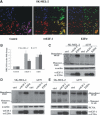
Co-localization of DREAM with wtE2F-1 or E2Ftr in the nuclei and homodimerization of DREAM after wtE2F-1 and E2Ftr overexpression. (A) After 24 hrs of infection in SK-MEL-2 cells, cells were incubated with DREAM antibody followed by Alexa-594 second antibody (shown in red, lower left panel) and E2F-1 antibody followed by Alexa-488 second antibody (shown in green, upper right panel) and then counterstained with DAPI (blue, upper left panel). An overlay (lower right panel) of yellow shows the co-localization of wtE2F-1 or E2Ftr with DREAM in the nuclei (magnification, ×600). Insets represent higher magnifications of the boxed areas (bar = 20 μm). (B) After 24 hrs of infection in SK-MEL-2 and A375 cells, RT-PCR of the DREAM gene was performed as described in Materials and methods. The bar graph is expressed as fold increase relative to control vector-infected cells adjusted for α-actin as an internal control. Each experiment is a representation of three independent experiments performed in duplicate (bars: mean ± S.D.). After 24 hrs of infection in SK-MEL-2 and A375 cells, 60 μg of total proteins from the whole cell lysates (C), cytoplasma (D) and nuclear (E) protein were extracted and subjected to Western blot analysis by using DREAM antibody. α-Actin was used for loading control of the whole cell lysates and cytoplasma fraction. PCNA was used for the loading control of the nuclear fraction (Ctrl: control; arrows: DREAM monomer or homodimer).
Fig 7
Fig 7
Schematic representation of the differences between the mechanism of wtE2F-1- and E2Ftr-induced apoptosis.

Similar articles

Cited by

References

    1. DeGregori J, Leone G, Miron A, et al. Distinct roles for E2F proteins in cell growth control and apoptosis. Proc Natl Acad Sci USA. 1997;94:7245–50. - PMC - PubMed
    1. Phillips AC, Vousden KH. E2F-1 induced apoptosis. Apoptosis. 2001;6:173–82. - PubMed
    1. Muller H, Bracken AP, Vernell R, et al. E2Fs regulate the expression of genes involved in differentiation, development, proliferation and apoptosis. Genes Dev. 2001;15:267–85. - PMC - PubMed
    1. Crosby ME, Almasan A. Opposing roles of E2Fs in cell proliferation and death. Cancer Biol Ther. 2004;3:1208–11. - PMC - PubMed
    1. Bandara LR, Buck VM, Zamanian M, et al. Functional synergy between DP-1 and E2F-1 in the cell cycle-regulating transcription factor DRTF1/E2F. EMBO J. 1993;12:4317–24. - PMC - PubMed

MeSH terms