. 2011 May 13;88(5):574-85.
doi: 10.1016/j.ajhg.2011.04.013.

Gain-of-function mutations of ARHGAP31, a Cdc42/Rac1 GTPase regulator, cause syndromic cutis aplasia and limb anomalies

Affiliations

Gain-of-function mutations of ARHGAP31, a Cdc42/Rac1 GTPase regulator, cause syndromic cutis aplasia and limb anomalies

Laura Southgate et al. Am J Hum Genet. .

Abstract

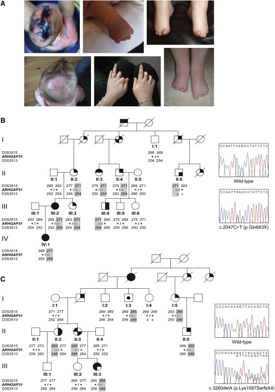
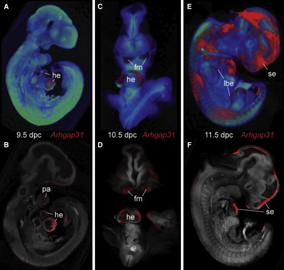
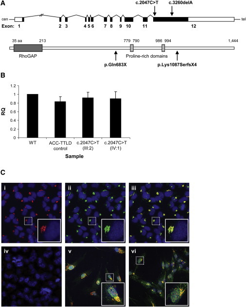
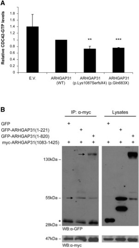
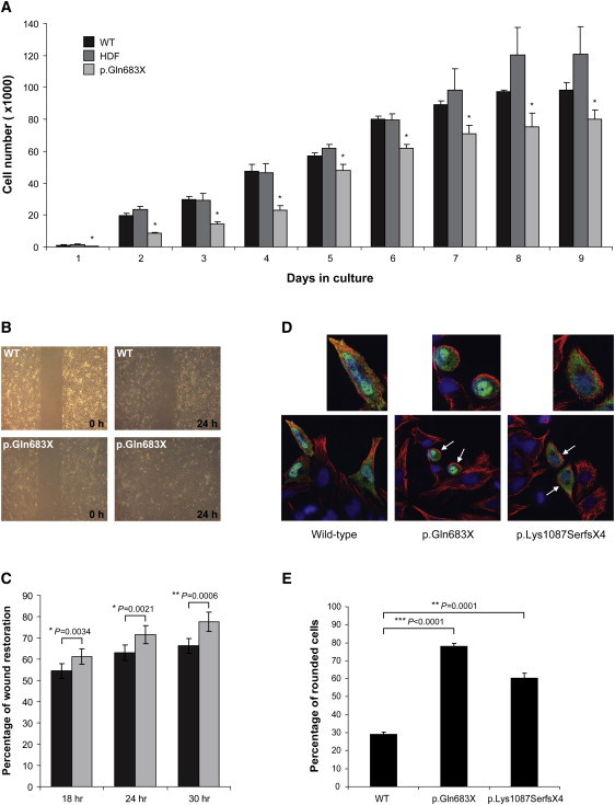
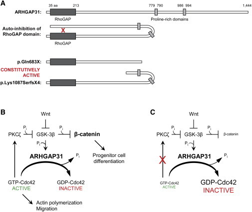
Regulation of cell proliferation and motility is essential for normal development. The Rho family of GTPases plays a critical role in the control of cell polarity and migration by effecting the cytoskeleton, membrane trafficking, and cell adhesion. We investigated a recognized developmental disorder, Adams-Oliver syndrome (AOS), characterized by the combination of aplasia cutis congenita (ACC) and terminal transverse limb defects (TTLD). Through a genome-wide linkage analysis, we detected a locus for autosomal-dominant ACC-TTLD on 3q generating a maximum LOD score of 4.93 at marker rs1464311. Candidate-gene- and exome-based sequencing led to the identification of independent premature truncating mutations in the terminal exon of the Rho GTPase-activating protein 31 gene, ARHGAP31, which encodes a Cdc42/Rac1 regulatory protein. Mutant transcripts are stable and increase ARHGAP31 activity in vitro through a gain-of-function mechanism. Constitutively active ARHGAP31 mutations result in a loss of available active Cdc42 and consequently disrupt actin cytoskeletal structures. Arhgap31 expression in the mouse is substantially restricted to the terminal limb buds and craniofacial processes during early development; these locations closely mirror the sites of impaired organogenesis that characterize this syndrome. These data identify the requirement for regulated Cdc42 and/or Rac1 signaling processes during early human development.


Figures

Figure 1
Figure 1
Features of ACC-TTLD and Segregation of ARHGAP31 Mutations (A) Characteristic phenotype of ACC-TTLD showing severe ACC (left panels) and a range of TTLD defects of the hands (middle panels) and feet (right panels), including partial absence of the fingers and toes and short distal phalanxes of fingers and toes. (B) Pedigree structure of family AOS-12 showing segregation of the c.2047C>T nonsense mutation represented in the adjacent sequence chromatogram. (C) Segregation and sequence chromatogram of the c.3260delA frameshift mutation in family AOS-5. Mutation carriers are denoted by +/–. Key to symbols: square, male; circle, female; upper left shading, aplasia cutis congenita; lower left shading, bony defect/abnormal fontanelle; upper right shading, terminal transverse limb defects; lower right shading, syndactyly; center shading, unsymptomatic mutation carrier; blank, unaffected.
Figure 2
Figure 2
Expression of Arhgap31 during Mouse Embryogenesis (A) Right lateral view of volume rendered OPT 3D representation of a 9.5 dpc mouse embryo showing Arhgap31 expression (in red) in developing heart (he). (B) Digital section of same embryo as in (A) showing expression in ventral wall of the early ventricle and atrium of the heart and the first pharyngeal arch (pa). (C) Frontal view of rendered and (D) digital coronal section of 10.5 dpc mouse embryo with expression in the lateral walls of the early ventricles of the heart and the first-pharyngeal-arch-derived facial mesenchyme (fm). (E and F) By 11.5 dpc the expression of Arhgap31 is restricted to distinct regions of the surface ectoderm (se), including the upper and lower limb bud (lbe).
Figure 3
Figure 3
Transcript and Protein Expression in WT and Mutant Cells (A) Schematic of the ARHGAP31 structure showing the position of the mutations identified in exon 12. The ARHGAP31 structure beneath depicts the known RhoGAP and proline-rich domains, a site of phosphorylation by GSK-3. (B) Real-time quantitative RT-PCR is used for the examination of ARHGAP31 transcript levels in lymphoblasts from two related subjects heterozygous for the c.2047C>T nonsense mutation as compared to a genotypically normal control (WT). Patient and control samples show no appreciable difference in transcript expression. Sample identifiers refer to the pedigree structure in Figure 1B. The ACC-TTLD control is a patient with no ARHGAP31 mutation (molecular genetic basis unknown). Data represent mean ± standard deviation (SD) from three independent experiments. RQ is used as an abbreviation for relative quantification. (C) Immunostaining of (i) endogenous ARHGAP31 (red) and (ii) Golgi (green); marked levels of colocalization to the Golgi apparatus in HeLa cells are visible (iii). The nucleus is stained in blue. (iv) The high specificity of the ARHGAP31 antibody is indicated by the absence of staining in the presence of blocking peptide to the binding epitope. In both (v) WT and (vi) mutant (p.Gln683X) fibroblasts, ARHGAP31 (green) localizes to the Golgi (red) and appears identical and of equivalent intensity. Images in the inset boxes show a 3× magnification of the single cells marked by the dashed lines.
Figure 4
Figure 4
Analysis of GAP Activity in ARHGAP31 Truncations (A) G-LISA assays measuring the relative amounts of Cdc42-GTP levels in HEK293 cells expressing Myc-tagged WT ARHGAP31 (full-length), p.Lys1087SerfsX4 or p.Gln683X. Relative Cdc42-GTP values are expressed as a ratio of Cdc42-GTP levels found in full-length ARHGAP31. Data are presented as mean ± SEM from four independent experiments. E.V. = empty vector; ∗∗p < 0.0002, ∗∗∗p < 0.00001. (B) Immunoprecipitation of mouse ARHGAP31 deletion constructs were used to map the intramolecular interaction between C-terminal amino acids 1083-1425 and the proximal 221 residues harboring the RhoGAP domain. Full-length protein products are marked by the arrows (smaller bands represent degradation products; the asterisk [] indicates the IgG light chain). Levels of transfected proteins, assessed by immunoblotting of the lysates with Myc antibody, are displayed in the lower panel.
Figure 5
Figure 5
Functional Characterization of ARHGAP31 Mutations (A) Bar chart comparing the proliferative activity of p.Gln683X primary dermal fibroblasts (light gray bars) with two distinct control fibroblast cell lines (black and dark gray bars). Data represent mean ± SEM for three independent experiments. Statistical analysis of each time point (days 1–9) revealed a significant decrease in the proliferative ability of cells carrying the p.Gln683X mutation when compared independently to each of the two unaffected controls (p < 0.01). The abbreviation WT indicates primary dermal fibroblasts from a tissue biopsy; HDF is used as an abbreviation for human dermal neonatal fibroblasts. (B) Wound-healing migration assay showing coverage of a cell-free gap by primary dermal fibroblasts heterozygous for the p.Gln683X mutation and WT control fibroblasts at 24 hr after wounding. (C) Plot showing percentage of wound restoration at 18, 24, and 30 hr after wounding. Fibroblasts heterozygous for the p.Gln683X mutation (light gray bars) migrate at a significantly faster rate than similar WT control fibroblasts (black bars). Data show mean ± SEM from three independent experiments. (D) HeLa cells transiently transfected with Myc-tagged WT ARHGAP31, p.Gln683X, and p.Lys1087SerfsX4 constructs. Cell shape was visualized by confocal microscopy for tubulin (red) and transfected cells identified by costaining with fluorescent conjugated Myc antibody (green). DAPI was used to stain the cell nuclei (blue). Rounded cells are indicated by the white arrows and 2× higher magnifications of individual cells are shown above. (E) Bar chart showing the mean percentage of rounded HeLa cells observed for each ARHGAP31 construct from three independent transfection experiments (error bars denote SD).
Figure 6
Figure 6
Schematic of Disrupted ARHGAP31 Signaling (A) Schematic representing the putative mechanism of disease. The C terminus of ARHGAP31 inhibits the activity of the RhoGAP domain by specific interaction with amino acids 1–221 (red “X”). Truncated mutant proteins lacking the C terminus would therefore be incapable of autoinhibition, and this would result in a constitutively active RhoGAP domain. (B) Schematic of the normal ARHGAP31 signaling system. ARHGAP31 cycles Cdc42 from an active to an inactive form by hydrolysis of GTP to GDP. GSK-3β upregulates ARHGAP31 levels, probably through phosphorylation at a consensus ERK1 site. Activated Cdc42 promotes actin polymerization and cellular processes, including migration, and acts to inhibit GSK-3β activity by stimulating PKCζ phosphorylation of GSK-3β. Wnt signaling is an additional negative regulator of GSK-3β activity. Downregulation of GSK-3β leads to decreased proteasomal degradation of cytosolic β-catenin. Active β-catenin translocates to the nucleus, whereupon the engagement of transcriptional cofactors controls the differentiation of progenitor cells in the skin. (C) In ACC-TTLD mutant cells, constitutive activation of ARHGAP31 leads to an imbalance between active and inactive Cdc42. A decrease in the levels of active GTP-bound Cdc42 results in reduced activation of PKCζ with a concomitant increase in β-catenin degradation and disruption of cellular processes. The following abbreviations are used: PKCζ, protein kinase C; GSK-3β, glycogen synthase kinase 3 beta; Pi, inorganic phosphate.

References

    1. Tcherkezian J., Lamarche-Vane N. Current knowledge of the large RhoGAP family of proteins. Biol. Cell. 2007;99:67–86. - PubMed
    1. Vega F.M., Ridley A.J. Rho GTPases in cancer cell biology. FEBS Lett. 2008;582:2093–2101. - PubMed
    1. Snape K.M., Ruddy D., Zenker M., Wuyts W., Whiteford M., Johnson D., Lam W., Trembath R.C. The spectra of clinical phenotypes in aplasia cutis congenita and terminal transverse limb defects. Am. J. Med. Genet. A. 2009;149A:1860–1881. - PubMed
    1. Tcherkezian J., Triki I., Stenne R., Danek E.I., Lamarche-Vane N. The human orthologue of CdGAP is a phosphoprotein and a GTPase-activating protein for Cdc42 and Rac1 but not RhoA. Biol. Cell. 2006;98:445–456. - PubMed
    1. Verdyck P., Blaumeiser B., Holder-Espinasse M., Van Hul W., Wuyts W. Adams-Oliver syndrome: Clinical description of a four-generation family and exclusion of five candidate genes. Clin. Genet. 2006;69:86–92. - PubMed

Publication types

MeSH terms

Supplementary concepts