. 2011 May 24;21(10):815-24.
doi: 10.1016/j.cub.2011.04.019. Epub 2011 May 12.

KIF4 regulates midzone length during cytokinesis

Affiliations

KIF4 regulates midzone length during cytokinesis

Chi-Kuo Hu et al. Curr Biol. .

Abstract

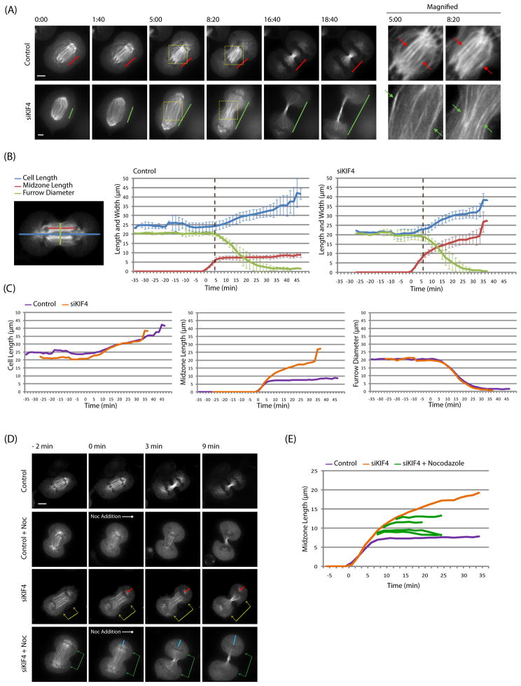
Background: Midzones, also called central spindles, are an array of antiparallel microtubules that form during cytokinesis between the separated chromosomes. Midzones can be considered to be platforms that recruit specific proteins and orchestrate cytokinetic events, such as sister nuclei being kept apart, furrow ingression, and abscission. Despite this important role, many aspects of midzone biology remain unknown, including the dynamic organization of midzone microtubules. Investigating midzone microtubule dynamics has been difficult in part because their plus ends are interdigitated and buried in a dense matrix, making them difficult to observe.

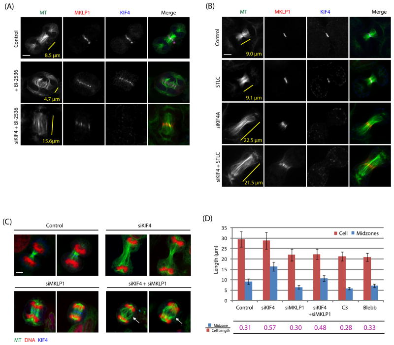
Result: We employed monopolar cytokinesis to reveal that midzone plus ends appear to be nondynamic. We identified the chromokinesin KIF4 as a negative regulator of midzone plus-end dynamics whose activity controls midzone length but not stability. KIF4 is required to terminate midzone elongation in late anaphase. In the absence of KIF4, midzones elongate abnormally, and their overlap regions are unfocused. Electron-dense material and midbodies are both absent from the elongated midzones, and actin filaments from the furrow cortex are not disassembled after ingression.

Conclusion: KIF4-mediated midzone length regulation appears to occur by terminating midzone elongation at a specific time during cytokinesis, making midzones and mitotic spindles differ in their dynamics and length-regulating mechanisms.


Figures

Figure 1
Figure 1. KIF4 negatively regulates midzone plus-end dynamics during monopolar cytokinesis
(A) Representative images of different stages in monopolar cytokinesis – polarization, cortex elongation/midzone termination, and furrow ingression. Cells were fixed and stained with antibodies as labeled. (B) Summary of monopolar cytokinesis phenotypes after depleting cytokinesis proteins. Depletions of PRC1, MKLP1, RacGAP1, and Ect2 blocked the initial polarization. KIF4 depleted midzones polarized, but were not terminated during monopolar cytokinesis. (C) KIF4 was required to terminate midzone plus-end dynamics in monopolar cytokinesis. HeLa cells stably expressing GFP-β-tubulin were imaged using spinning disk confocal microscope (Movie 1, 2). Imaging started at time zero. Monopolar midzones plus-ends were not polymerizing during furrow ingression in control (red arrow), but were growing towards the furrow region in KIF4 depleted cells (green arrow). (D) KIF4 was required to properly localize proteins on monopolar midzones. KIF4 depleted monopolar midzone polymerized into the furrow-like cortex region, which was marked in dotted yellow line according to Anillin localization on the cortex. Furrow regions were boxed and magnified in the right. PRC1, MKLP1, and Aurora B localized differently in the absence of KIF4 during monopolar cytokinesis. Bars = 5 μm. See also Figure S1.
Figure 2
Figure 2. KIF4 mediated plus-end termination regulates midzone length in bipolar cytokinesis
(A) KIF4 regulated constant midzone lengths during normal bipolar cytokinesis. HeLa cells expressing GFP-β-tubulin were imaged using spinning disk confocal microscope (Movie 5, 6). Time zero was the point furrow ingression started. Midzone length was defined as the distance between segregated chromosomes. Note that midzone lengths were constant in control (red lines), but increased with time in KIF4 depleted cell (green lines) throughout furrow ingression. Antiparallel microtubule overlapping regions were boxed and magnified in the right. The overlaps were constant in length in controls (red arrows) but not in KIF4 depleted cells (green arrows). (B, C) Correlations between cortex elongation, midzone elongation, and furrow ingression in normal and KIF4 depleted cells. Twenty controls and ten KIF4 depleted cells stably expressing GFP-β-tubulin were measured, averaged, plotted, and grouped differently to facilitate the comparisons. Definition of cell length, midzone length, and furrow diameter were shown. Time zero referred to the point chromosome segregation started (anaphase onset). Error bars in (B) were standard deviations and were omitted in (C) for clarity. Coincident with furrow initiation (dotted brown line), anaphase midzones elongation was terminated in controls, but not in KIF4 depleted cells. Velocities of midzones elongation in KIF4 depleted cells were similar before and after furrow ingression (3.83 and 3.47 μm/min). The velocities of cortex elongation and furrow ingression were similar between controls and KIF4 depleted cells. (D) KIF4 depleted midzones were resistant to 10 μM nocodazole. HeLa cells expressing GFP-β-tubulin were imaged using spinning disk confocal microscope (Movie 8). Midzone length was defined as the distance between segregated chromosomes and nocodazole was added at time zero. KIF4 depleted midzone continuously elongated after furrow ingression (yellow arrows) which pushed chromosomes along with elongated cortex (red lines). Nocodazole stopped the continuous elongation of KIF4 depleted midzones (green arrows). The distance between chromosomes and the polar cortex increased while cortex elongation continued (blue lines). (E) Nocodazole stopped post-furrow elongation of KIF4 depleted midzones. Midzone lengths of four KIF4 depleted cells treated with 10 μM nocodazole after furrow ingression were measured, plotted, and superimposed with the midzone length plot in (C) by their lengths before nocodazole exposure. KIF4 depleted midzones were constant in length after nocodazole treatment. Bars = 5 μm. Also see Figure S2.
Figure 3
Figure 3. KIF4 mediated midzone termination focuses midzone proteins on midzones
(A) Midzone proteins relocalized differently on KIF4 depleted midzones. Cells were fixed and co-stained as labeled. CENPE was used as a reference for protein localization comparison. In the absence of KIF4, Aurora B preferentially relocalized to the furrow cortex. Plk1 and MKLP1 to be less diffuse, and PRC1 diffuse all along midzone microtubules as CENPE did. (B) Experiment design to replace endogenous KIF4 with mouse KIF4 using human KIF4 specific siRNA. (C) Summary of expressed mouse GFP-KIF4 (mKIF4). (D) GFP-mKIF4-FL and GFP-mKIF4-tailless focused and colocalized with PRC1 on endogenous KIF4 depleted midzones. GFP-mKIF4-headless did not focus on midzones and could not be rescued by endogenous KIF4. PRC1 focusing was rescued by GFP-mKIF4-FL, tailless, but not headless. Bars = 5 μm. Also see Figure S3.
Figure 4
Figure 4. KIF4 mediated midzone termination focuses furrow proteins and disassembles actin filaments on the cortex
(A) KIF4 was required to focus furrow on the cortex during cytokinesis. Cells at different stages were fixed and stained with Anillin as a furrow marker. Furrow regions were focusing with time in controls, but not in KIF4 depleted cells. Bars = 5 μm. (B) Lengths of cell and furrow region in (A) were measured and plotted. Definitions of cell length and furrow region were shown. Data were fitted with exponential trend lines. Furrow regions were focusing while cell length were increasing in controls (purple dots and line), but not in KIF4 depleted cells (orange dots and line). Cell numbers in control = 25 and in siKIF4 = 50. (C) Thin section EM of furrow regions in cytokinesis. KIF4 depletion caused an elongated intracellular bridge between two daughter cells. Microtubules (green arrows) were less bundled and in lower density in KIF4 depleted cells. Electron dense materials, which coated central overlaps of midzones in controls (brown arrows), was missing in KIF4 depleted cells. The whole-cell views were shown as insets. Boxed regions were magnified in the right. (D) Thin section EM of the furrow cortex. Long straight actin filaments (red arrows) were observed underneath the cortex in the unfocused furrow region of KIF4 depleted cells. Electron dense materials (white arrows) were observed coating on actin filaments but not on microtubules (green arrow). Also see Figure S4.
Figure 5
Figure 5. KIF4 mediated termination of midzone elongation regulates midzone length in cytokinesis
(A) KIF4 depletion overrode BI-2536-caused block of midzone elongation. Midzone length was shorter than controls under BI-2536 treatment, but became longer than controls if KIF4 was further depleted. Midzone length was measured by the distance between chromosomes. (B) Kinesin-5 inhibitor STLC did not block midzone elongation. KIF4 depleted midzones were longer than controls and in similar lengths with or without STLC treatment. (C–D) Centralspindlin was not involved in midzone elongation. Compared to controls, MKLP1 depletion caused shorter cell length and midzones, but maintained a similar midzone over cell length ratio, unless KIF4 was absent. RhoA inhibitor C3 and Myosin II inhibitor blebbistatin (Blebb), which block the cortex elongation, also caused comparable ratios. Error bars were standard deviations. Cell numbers in control = 41, in siKIF4 = 41, in siMLP1 = 41, in siKIF4/siMKLP1 = 26, in C3 = 41, and in Blebb = 68.
Figure 6
Figure 6. Model of KIF4 mediated midzone plus-end termination regulates midzone length in cytokinesis
Different to mitotic spindles regulating its length through the balance between dynamic plus-ends growing and shrinking, and motor mediated microtubule sliding, midzones regulate its length through a KIF4 mediated termination of anaphase midzone elongation. Negatively regulated by PLk1, KIF4 terminates midzone elongation by inhibiting microtubule plus-end polymerization. A microtubule (MT) stabilizer, which inhibits plus-end depolymerization, provides midzone stability.

Similar articles

Cited by

References

    1. Glotzer M. The 3Ms of central spindle assembly: microtubules, motors and MAPs. Nat Rev Mol Cell Biol. 2009;10:9–20. - PMC - PubMed
    1. Gruneberg U, Neef R, Li X, Chan EH, Chalamalasetty RB, Nigg EA, Barr FA. KIF14 and citron kinase act together to promote efficient cytokinesis. J Cell Biol. 2006;172:363–372. - PMC - PubMed
    1. Bringmann H. Cytokinesis and the spindle midzone. Cell Cycle. 2005;4:1709–1712. - PubMed
    1. Euteneuer U, McIntosh JR. Polarity of midbody and phragmoplast microtubules. J Cell Biol. 1980;87:509–515. - PMC - PubMed
    1. Brust-Mascher I, Civelekoglu-Scholey G, Kwon M, Mogilner A, Scholey JM. Model for anaphase B: role of three mitotic motors in a switch from poleward flux to spindle elongation. Proc Natl Acad Sci U S A. 2004;101:15938–15943. - PMC - PubMed

Publication types

LinkOut - more resources