. 2011 Jun;13(6):635-48.
doi: 10.1093/neuonc/nor021. Epub 2011 May 12.

Local interstitial delivery of z-butylidenephthalide by polymer wafers against malignant human gliomas

Affiliations

Local interstitial delivery of z-butylidenephthalide by polymer wafers against malignant human gliomas

Horng-Jyh Harn et al. Neuro Oncol. 2011 Jun.

Abstract

We have shown that the natural compound z-butylidenephthalide (Bdph), isolated from the chloroform extract of Angelica sinensis, has antitumor effects. Because of the limitation of the blood-brain barrier, the Bdph dosage required for treatment of glioma is relatively high. To solve this problem, we developed a local-release system with Bdph incorporated into a biodegradable polyanhydride material, p(CPP-SA; Bdph-Wafer), and investigated its antitumor effects. On the basis of in vitro release kinetics, we demonstrated that the Bdph-Wafer released 50% of the available Bdph by the sixth day, and the release reached a plateau phase (90% of Bdph) by the 30th day. To investigate the in situ antitumor effects of the Bdph-Wafer on glioblastoma multiforme (GBM), we used 2 xenograft animal models-F344 rats (for rat GBM) and nude mice (for human GBM)-which were injected with RG2 and DBTRG-05MG cells, respectively, for tumor formation and subsequently treated subcutaneously with Bdph-Wafers. We observed a significant inhibitory effect on tumor growth, with no significant adverse effects on the rodents. Moreover, we demonstrated that the antitumor effect of Bdph on RG2 cells was via the PKC pathway, which upregulated Nurr77 and promoted its translocation from the nucleus to the cytoplasm. Finally, to study the effect of the interstitial administration of Bdph in cranial brain tumor, Bdph-Wafers were surgically placed in FGF-SV40 transgenic mice. Our Bdph-Wafer significantly reduced tumor size in a dose-dependent manner. In summary, our study showed that p(CPP-SA) containing Bdph delivered a sufficient concentration of Bdph to the tumor site and effectively inhibited the tumor growth in the glioma.


Figures
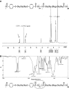
Fig. 1.
Fig. 1.
Structural analysis of the CPP-SA copolymer. (A) 1H NMR spectroscopy of the CPP-SA copolymer. The characteristic signals of aromatic protons in CPP were observed at 6.9–8.2 ppm. Another characteristic signal of methylene protons in SA was measured at 1.3 ppm. (B) Fourier transform infrared spectroscopy of the CPP-SA copolymer. The characteristic signal of the anhydride bond between CPP and SA (shown circled in red) was observed at 1812.76 cm−1.
Fig. 2.
Fig. 2.
The release kinetics of 10% and 15% Bdph-Wafers measured over 30 days in vitro. Sustained release of Bdph was observed with both wafer formulations. *P < .05. n = 3 for each concentration.
Fig. 3.
Fig. 3.
Growth inhibition of rat malignant glioma cells by Bdph-Wafers. (A) The rat malignant glioma cell line RG2 was treated with control wafers and with 3% and 10% Bdph-Wafers for 24 h. Cell viability was determined with the 3-[4,5-dimethylthiazol-2-yl]-2,5-diphenyltetrazolium bromide (MTT; Sigma) assay. Data shown are the mean ± standard deviation from 3 independent experiments. **P < .01. (B) Cell morphology of RG2 malignant glioma cells treated for 24 h with control wafers and with 3% and 10% Bdph-Wafers.
Fig. 4.
Fig. 4.
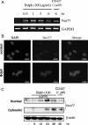
Induction of Nur77 transcripts and migrate from the nucleus to the cytoplasm after Bdph exposure. (A) Rat glioblastoma multiforme (GBM) cells (RG2) were treated with Bdph (100 µg/mL) for the indicated time periods (0, 0.5, 1, 3, and 6 h). CD437 (1 µM) was used as the Nur77-positive control. After incubation with drugs, cells were collected, and total RNA was isolated. Glyceraldehyde-3-phosphate dehydrogenase (GAPDH) was used as an internal control. (B) Rat GBM cells (RG2) cells were treated with Bdph (100 µg/mL) for 24 h and then immunostained with Nur77 antibody followed by the corresponding rhodamine-conjugated anti-immunoglobulin G secondary antibody to show Nur77 protein localization. Nuclei were stained with 4′-6-diamidino-2-phenylindole (DAPI). Staining was visualized with a fluorescence microscope (bar = 50 µm). (C) Bdph-induced Nur77 translocation from the nucleus to the cytoplasm in RG2 cells. RG2 cells were plated in 10-cm dishes and incubated until 90% confluent. Cells were treated with Bdph (100 μg/mL) for different time periods (0, 6, 12, 24, and 48 h). The cells were harvested, and nuclear and cytoplasmic fractions were isolated. CD437 (1 µM) was used as the Nur77-positive control. Nur77 expression in cytoplasmic and nuclear fractions was evaluated by Western blot. β-actin was used as an internal control.
Fig. 5.
Fig. 5.
Role of signaling pathways in Bdph-induced growth inhibition. (A) RG2 cells were treated with Bdph (100 μg/mL) for 0 to 180 min as indicated. Western blot analysis was performed with pPKC, pJNK, JNK, pERK, ERK, pAKT, and AKT antibodies. Expression of β-actin was used as an internal control. (B) RG2 cells were pretreated with a PKC inhibitor Go6983 (0.5−1 μmol/L), JNK inhibitor SP600125 (5−10 μmol/L), mitogen-activated protein kinase kinase 1/2 inhibitor PD98059 (5−10 μmol/L), or the PI3K/AKT/GSK3β inhibitor wortmannin (20–40 μmol/L) for 1 h. Subsequently, the RG2 cells were treated with 100 μg/mL Bdph for 24 h. The viability was determined with a 3-[4,5-dimethylthiazol-2-yl]-2,5- diphenyltetrazolium bromide (MTT; Sigma) assay. *P < .05. (C) PKCδ signaling pathways involved in the Bdph-induced Nur77 migration. RG2 Cells were treated with Bdph (100 μg/mL) for 1 h. Western blot analysis was performed for pPKC (pan, α, β, δ, θ, ξ, λ) antibodies. (D) RG2 cells were pretreated with PKCδ siRNA (50 and 100 nM) for 1 h and subsequently treated with Bdph (100 µg/mL) for 1 h. Total protein was extracted after treatment. pPKCδ expression level was analyzed by Western blotting. (E) RG2 cells were pretreated with PKCδ siRNA (50 and 100 nM) for 1 h and were subsequently treated with Bdph (100 µg/mL) for 24 h. Nur77 expression in cytoplasm and nucleus fractions was evaluated by Western blot. Expression of β-actin was used as an internal control.
Fig. 6.
Fig. 6.
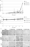
Bdph-Wafers inhibited tumor growth in a syngeneic rat glioblastoma multiforme (GBM) model. RG2 cells (1 × 10⁶) were implanted subcutaneously into the hind-flank region of F344 rats. Five days after RG2 cell transplantation, the rats were treated with wafers alone, 3% Bdph-Wafers, 10% Bdph-Wafers, or 3% BCNU-Wafers. The resulting tumor sizes (A) and rat body weights (B) are shown as the mean ± standard deviation *P < .05. (C) Immunohistochemical staining was performed on GBM tissues (at day 30 after implantation of RG2 cells). Sections of GBM tumors from animals receiving wafers only, 3% Bdph-Wafers, 10% Bdph-Wafers, and 3% BCNU-Wafers were immunohistochemically stained for Ki-67, cleaved caspase-3, or Nur77. Positive cells are stained brown (original magnification, ×400; bar = 50 µm).
Fig. 7.
Fig. 7.
Bdph-Wafers inhibited tumor growth in a human glioblastoma multiforme (GBM) xenograft nude mouse model. DBTRG-05MG cells (1 × 10⁶) were implanted subcutaneously into the hind-flank region of Foxn1 nu/nu mice. Five days after transplantation, mice were treated with wafers only or with 3% and 10% Bdph-Wafers. Tumor sizes (A) and body weights (B) are shown as the mean ± standard deviation *P < .05. **P < .01 (C) Tumors isolated from animals treated with wafers only or with 3% and 10% Bdph-Wafers.
Fig. 8.
Fig. 8.
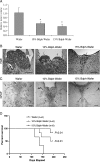
Bdph-Wafers inhibited tumor growth and survival study in a spontaneous brain tumor mouse model. FGF-SV40 transgenic mice were implanted with control or 10% or 15% Bdph-Wafers to verify antitumor activity of Bdph. Thirty days after wafer implantation, the mice were killed, and the tumors were analyzed. (A) The relative tumor area ± standard deviation is shown for each group (*P < .05). (B) Hematoxylin and eosin staining of coronal sections (original magnification, ×200; bar = 100 µm). (C) Immunohistochemical staining for GFAP. Positive cells are stained brown (original magnification, ×100; bar = 100 µm). Arrow head indicates the area of tumor. (D) Ten percent and 15% Bdph wafer, as well as 15% BCNU wafer, were implanted into FGF-1-SV40 transgeneic mice, and survival was studied for 200 days. Mice that were implanted with 15% BCNU died within two days. By using Kaplan-Meier survival curves analysis, the relative survival rate is shown for each group (*P < .05). The 15% Bdph wafer had the best survival rate after 200 days. The survival rate depended on Bdph concentration.

References

    1. Santarius T, Kirsch M, Rossi ML, Black PM. Molecular aspects of neuro-oncology. Clin Neurol Neurosur. 1997;99:184–195. doi:10.1016/S0303-8467(97)00025-5. - DOI - PubMed
    1. Cobb MA, Husain M, Andersen BJ, al-Mefty O. Significance of proliferating cell nuclear antigen in predicting recurrence of intracranial meningioma. J Neurosurg. 1996;84:85–90. doi:10.3171/jns.1996.84.1.0085. - DOI - PubMed
    1. Giese A, Bjerkvig R, Berens ME, Westphal M. Cost of migration: invasion of malignant gliomas and implications for treatment. J Clin Oncol. 2003;21:1624–1636. doi:10.1200/JCO.2003.05.063. - DOI - PubMed
    1. Blacklock JB, Wright DC, Dedrick RL, et al. Drug streaming during intra-arterial chemotherapy. J Neurosurg. 1986;64:284–291. doi:10.3171/jns.1986.64.2.0284. - DOI - PubMed
    1. Shapiro WR, Green SB. Reevaluating the efficacy of intra-arterial BCNU. J Neurosurg. 1987;66:313–315. - PubMed

Publication types

MeSH terms