. 2011 Jul 1;17(13):4309-19.
doi: 10.1158/1078-0432.CCR-10-1820. Epub 2011 May 12.

MAGE-A inhibits apoptosis in proliferating myeloma cells through repression of Bax and maintenance of survivin

Affiliations

MAGE-A inhibits apoptosis in proliferating myeloma cells through repression of Bax and maintenance of survivin

Tricia Nardiello et al. Clin Cancer Res. .

Abstract

Purpose: The type I Melanoma Antigen GEnes (MAGEs) are commonly expressed in cancers, fueling speculation that they may be therapeutic targets with oncogenic potential. They form complexes with RING domain proteins that have E3 ubiquitin ligase activity and promote p53 degradation. MAGE-A3 was detected in tumor specimens from patients with multiple myeloma and its expression correlated with higher frequencies of Ki-67(+) malignant cells. In this report, we examine the mechanistic role of MAGE-A in promoting survival of proliferating multiple myeloma cells.

Experimental design: The impact of MAGE-A3 expression on survival and proliferation in vivo was examined by immunohistochemical analysis in an independent set of tumor specimens segregated into two groups: newly diagnosed, untreated patients and patients who had relapsed after chemotherapy. The mechanisms of MAGE-A3 activity were investigated in vitro by silencing its expression by short hairpin RNA interference in myeloma cell lines and primary cells and assessing the resultant effects on proliferation and apoptosis.

Results: MAGE-A3 was detected in a significantly higher percentage of relapsed patients compared with newly diagnosed, establishing a novel correlation with progression of disease. Silencing of MAGE-A showed that it was dispensable for cell cycling, but was required for survival of proliferating myeloma cells. Loss of MAGE-A led to apoptosis mediated by p53-dependent activation of proapoptotic Bax expression and by reduction of survivin expression through both p53-dependent and -independent mechanisms.

Conclusions: These data support a role for MAGE-A in the pathogenesis and progression of multiple myeloma by inhibiting apoptosis in proliferating myeloma cells through two novel mechanisms.


Figures

Figure 1
Figure 1. Immunohistochemical analysis of CTAg expression in newly diagnosed and relapsed myeloma patients
A. Bone marrow biopsy sections were stained with a panel of mAb recognizing several CTAg (MAGE-A3 and CT7 shown). Representative sections are depicted. Brown chromophore indicates positive staining for CTAg (arrows). 20X magnification. B. Frequency of expression for each CTAg in new and relapsed patients were plotted and compared. C. Grading of MAGE-A3 expression (percent of MAGE-A3+ MM cells) was divided into quartiles (focal-25%, 25-50%, 50-74%, and 75-100%) and plotted by the percentage of patients in each quartile. D. Plasma Cell Proliferation Index was plotted for newly diagnosed and relapsed MM patient specimens. Mean PCPI ± standard error of the mean depicted. P-values calculated by Student's two-tailed t-test.
Figure 2
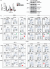
Figure 2. Silencing of MAGE-A does not directly affect proliferation in MM cells
MM.1r and ARP-1 HMCL and Pt #1 cells were transduced with MAGE-A-targeted (shMA 128375 and 129750) or non-target (shNT) shRNA lentiviral particles and samples were harvested at the indicated time points. A. MAGE-A3 mRNA in MM.1r cells was assessed by semi-quantitative real time (q) RT-PCR. Results were normalized to shNT control at each time point. Error bars depict standard error of the mean for triplicates of each sample. Control (Con), untreated negative control cells. B. HMCL and Pt #1 cells were treated with shMA 128375 or controls and MAGE-A3 protein expression was assessed by western blot at 48 hrs. Relative optical density (rel OD) was normalized to matched β-actin and expressed as a fraction of shNT control. C. shMA-transduced and control cells were pulsed for 30 minutes with 10 mM BrdU at the indicated time points. Proliferation was assessed by intracellular staining for BrdU and 7-AAD, followed by flow cytometry. Acquisition populations were gated on viable cells in forward vs. side scatter plots (Live Gate, insets) and the gated events plotted by 7-AAD (total DNA content) vs. FITC (BrdU incorporation) fluorescence, resulting in segregation into the G1, S, and G2/M populations as labeled in the first panel.
Figure 3
Figure 3. Silencing of MAGE-A results in activation of intrinsic apoptosis
HMCL and Pt #1 cells were transduced shMA or controls as previously described and apoptosis was assessed by staining with annexin V. A. Total (ungated) acquisition populations at 72 or 96 hrs were plotted by Annexin V-PE (apoptosis) vs. 7-AAD (necrosis) fluorescence. B. The percentages of viable cells (Live Gate as described in fig. 2, solid circles) and annexin V+ cells in the ungated acquisition population (solid squares) were plotted over time for the MAGE-A-silenced and control groups. C. HMCL and Pt#1 cells were stained with MitoTracker Red®, which is fixed in mitochondria with intact membrane polarization, and analyzed by flow cytometry. Total (ungated) events were plotted by MitoTracker Red® fluorescence and mitochondrial depolarization was assessed by decreased fluorescence (bars). E. Lentivirus-transduced cells were incubated with 10 μM Q-VD-OPh or DMSO vehicle control for 72 hrs and apoptosis was assessed by staining with annexin V. D. Caspase-3 and -9 were analyzed by western blot in at 48 hrs for MM.1r and Pt #1 and 72 hrs for ARP-1 cells.
Figure 4
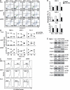
Figure 4. Silencing of MAGE-A induces p53-dependent Bax/Bak expression and repression of survivin
MM.1r cells were transduced with MAGE-A-targeted (shMA) or p53-targeted (shp53) shRNA lentiviral constructs, both targeted constructs, or controls as indicated and cells were harvested at 48 hrs. A. Bax, Bak, and survivin mRNA expression was assessed by qRT-PCR. B. Protein expression of Bcl-2 proteins and survivin was assessed by western blot. C. p53 mRNA expression was assessed by qRT-PCR and protein expression by western blot. D. Cells were incubated with 40 nM MG132 prior to harvest and lysates were immunoprecipitated (IP) with p53-specific mAb or isotype control mu IgG (iso). Ubiquitinylation of p53 was assessed by western blot.
Figure 5
Figure 5. Silencing of MAGE-A results in repression of survivin in the absence of p53
ARP-1 (p53-/-) and Pt #1 (p53mut/-) cells were transduced with MAGE-A-targeted (shMA) shRNA lentiviral constructs or controls as indicated and cells were harvested at 48 hrs (Pt #1) or 72 hrs (ARP-1). A. p53 protein in Pt #1 cells was assessed by western blot. B. Bax, Bak, and survivin mRNA expression was assessed by qRT-PCR. C. Protein expression of Bax, Bak, and survivin was assessed by western blot.

Similar articles

Cited by

References

    1. Simpson AJ, Caballero OL, Jungbluth A, Chen YT, Old LJ. Cancer/testis antigens, gametogenesis and cancer. Nat Rev Cancer. 2005;5:615–25. - PubMed
    1. Scanlan MJ, Gure AO, Jungbluth AA, Old LJ, Chen YT. Cancer/testis antigens: an expanding family of targets for cancer immunotherapy. Immunol Rev. 2002;188:22–32. - PubMed
    1. Jungbluth AA, Ely S, DiLiberto M, et al. The cancer-testis antigens CT7 (MAGE-C1) and MAGE-A3/6 are commonly expressed in multiple myeloma and correlate with plasma-cell proliferation. Blood. 2005;106:167–74. - PubMed
    1. Kyle RA, Rajkumar SV. Multiple myeloma. Blood. 2008;111:2962–72. - PMC - PubMed
    1. Monte M, Simonatto M, Peche LY, et al. MAGE-A tumor antigens target p53 transactivation function through histone deacetylase recruitment and confer resistance to chemotherapeutic agents. Proc Natl Acad Sci U S A. 2006;103:11160–5. - PMC - PubMed

Publication types

MeSH terms