. 2011 Jun;22(6):1137-43.
doi: 10.1681/ASN.2010090967. Epub 2011 May 12.

Anti-phospholipase A2 receptor antibody in membranous nephropathy

Affiliations

Anti-phospholipase A2 receptor antibody in membranous nephropathy

Weisong Qin et al. J Am Soc Nephrol. 2011 Jun.

Abstract

The M-type phospholipase A2 receptor (PLA2R) is a target autoantigen in adult idiopathic membranous nephropathy (MN), but the prevalence of autoantibodies against PLA2R is unknown among Chinese patients with MN. Here, we measured anti-PLA2R antibody in the serum of 60 patients with idiopathic MN, 20 with lupus-associated MN, 16 with hepatitis B (HBV)-associated MN, and 10 with tumor-associated MN. Among patients with idiopathic MN, 49 (82%) had detectable anti-PLA2R autoantibodies using a Western blot assay; an assay with greater sensitivity detected very low titers of anti-PLA2R in 10 of the remaining 11 patients. Using the standard assay, we detected anti-PLA2R antibody in only 1 patient with lupus, 1 with HBV, and 3 with cancer, producing an overall specificity of 89% in this cohort limited to patients with secondary MN. The enhanced assay detected low titers of anti-PLA2R in only 2 additional samples of HBV-associated MN. In summary, these results suggest that PLA2R is a major target antigen in Chinese idiopathic MN and that detection of anti-PLA2R is a sensitive test for idiopathic MN.


Figures

Figure 1.
Figure 1.
Western blot analysis of representative serum samples from patients with idiopathic MN demonstrates reactivity with native and recombinant PLA2R. Extracts of HGE and recombinant human PLA2R (rPLA2R) were electrophoresed under nonreducing conditions and immunoblotted with patient serum (IMN1 through IMN4) at 1:25 and detected with anti-human IgG4. IMN1, IMN3, and IMN4 recognize PLA2R in HGE and the smaller cell-expressed rPLA2R. The recognition was confirmed using commercial polyclonal anti-PLA2R antibody. IMN2 in this figure is negative. No other reactive bands were observed. M. Protein standard (kD).
Figure 2.
Figure 2.
Idiopathic MN sera react with glycosylated and deglycosylated PLA2R. Native PLA2R in HGE is heavily glycosylated and treatment with peptide N-glycosidase F (PNGaseF) caused a downward shift to approximately 145 kD. The anti-PLA2R antibodies from patients IMN1, IMN3, and IMN4 recognized the deglycosylated as well as the native PLA2R. M. Protein standard (kD).
Figure 3.
Figure 3.
Increasing the sensitivity of the western blot assay reveals low titers of anti-PLA2R in initially negative idiopathic MN sera. Representative examples of immunoblots of HGE with two idiopathic MN sera that were negative under standard conditions (IMN2 and IMN8) but became positive when the dilution of serum sample was reduced to 1:10 and the exposure time was prolonged to 10 minutes. PC is a MN serum that was positive under standard conditions and is diluted to 1:1000 for this experiment. The downward shift in the size of the reactive band could be observed after PNGaseF treatment. HC, healthy control, serum from healthy adult (1:10). M. Protein standard (kD).
Figure 4.
Figure 4.
The prevalence of anti-PLA2R in idiopathic MN (IMN) in remission is low by comparison to IMN patients with nephrotic syndrome. The prevalence of anti-PLA2R seropositivity was compared in the 60 patients with nephrotic syndrome to 21 additional patients in remission (see Supplemental Tables 2 and 3 for details), using the standard assay.
Figure 5.
Figure 5.
Anti-PLA2R is detected infrequently in secondary MN. The dark bars represent the prevalence of anti-PLA2R detected in patients with idiopathic (IMN), lupus-associated (LMN), hepatitis B—associated (HBV-MN), and cancer-associated (Ca-MN) membranous nephropathy, using the standard Western blot assay.
Figure 6.
Figure 6.
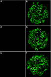
Staining for IgG4 deposition in glomeruli of secondary MN patients distinguishes those with circulating anti-PLA2R from anti-PLA2R-negative cases. (A,C,E) Negative immunostaining for IgG4 in glomeruli of patients with lupus-MN (A), HBV-MN (C), and tumor-associated MN (E) but without circulating anti-PLA2R antibodies. (B,D,F) Bright IgG4 immunostaining in glomeruli of patients with lupus-MN (B), HBV-MN (D), and tumor-associated MN (F) whose circulating anti-PLA2R antibodies were positive.

References

    1. Wasserstein AG: Membranous glomerulonephritis. J Am Soc Nephrol 8: 664–674, 1997 - PubMed
    1. Li LS, Liu ZH: Epidemiologic data of renal diseases from a single unit in China: Analysis based on 13,519 renal biopsies. Kidney Int 66: 920–923, 2004 - PubMed
    1. Kerjaschki D: Pathomechanisms and molecular basis of membranous nephropathy. Lancet 364: 1194–1196, 2004 - PubMed
    1. Salant DJ, Belok S, Madaio MP, Couser WG: A new role for complement in experimental membranous nephropathy in rats. J Clin Invest 66: 1339–1350, 1980 - PMC - PubMed
    1. Salant DJ: In search of the elusive membranous nephropathy antigen. Nephron Physiol 112: 11–12, 2009 - PubMed

Publication types

LinkOut - more resources