. 2011 Jul;25(7):1126-36.
doi: 10.1210/me.2010-0463. Epub 2011 May 12.

GATA4 regulates estrogen receptor-alpha-mediated osteoblast transcription

Affiliations

GATA4 regulates estrogen receptor-alpha-mediated osteoblast transcription

Gustavo A Miranda-Carboni et al. Mol Endocrinol. 2011 Jul.

Abstract

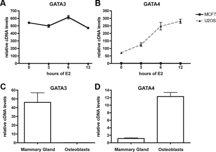
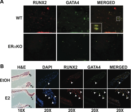
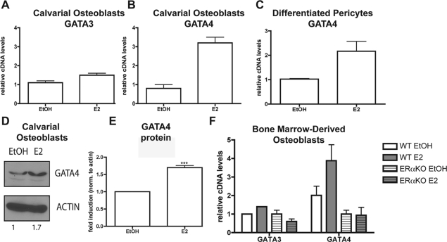
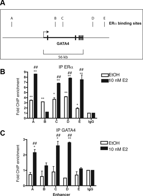
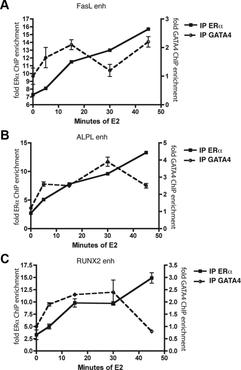
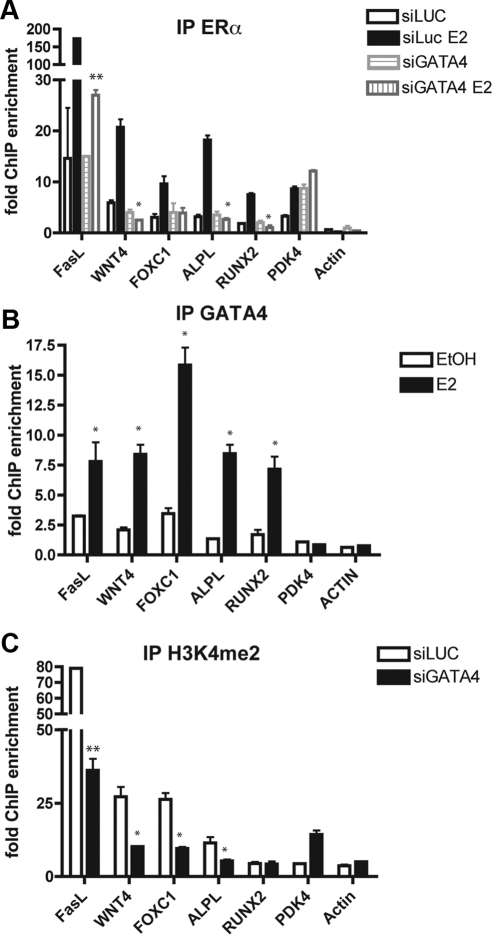
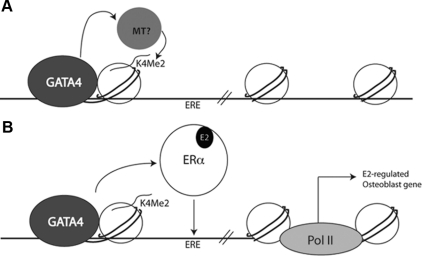
Estrogens regulate osteoblast differentiation and mineralization. We identified GATA4 as a transcription factor expressed in osteoblasts and directly regulated by 17β-estradiol in this cell type but not in breast cancer cells, another estrogen-responsive tissue. Chromatin immunoprecipitation sequencing (chromatin immunoprecipitation sequencing) reveals that estrogen receptor α (ERα) binds to chromatin near GATA4 at five different enhancers. GATA4 and ERα are both recruited to ERα binding sites near genes that are specifically expressed in osteoblasts and control osteoblast differentiation. Maximal binding of GATA4 precedes ERα binding, and GATA4 is necessary for histone 3 lysine 4 dimethylation at ERα binding sites, suggesting that GATA4 is a pioneer factor for ERα. As such, knockdown of GATA4 reduced recruitment of ERα to DNA. Our study illustrates that GATA4 is a pioneer factor for ERα recruitment to osteoblast-specific enhancers.


Figures

Fig. 1.
Fig. 1.
GATA4 is expressed in osteoblasts. A, U2OS-ERα and MCF-7 cells were deprived of estrogen for 3 d in phenol red-free media containing 5% CDT-FBS. They were then treated with 10 nm E2 for 0, 3, 6, or 12 h, and RNA was obtained. GATA3 mRNA was analyzed by qPCR and normalized to actin mRNA. B, Cells were treated as in A, and GATA4 mRNA was analyzed by qPCR and normalized to actin mRNA. C, Mammary gland and osteoblast mRNA was obtained and converted to cDNA. GATA3 mRNA was analyzed by qPCR and normalized to actin mRNA. D, Cells were treated as in C, and GATA4 mRNA was analyzed by qPCR and normalized to actin mRNA. Error bars in all panels represent ±1 sd.
Fig. 2.
Fig. 2.
GATA4 is localized to osteoblasts in vivo and is induced by E2. A, Femurs from wild-type (WT) and ERαKO mice were snap-frozen and embedded in OCT. The 10-μm sections were stained with RUNX2 and GATA4. Images are of trabecular bone near the growth plate. B, Calvariae from neonatal d-2 mice were grown as organ cultures for 7 d and then treated with vehicle control [ethanol (EtOH)] or 10 nm E2 for 24 h. Calvariae were fixed and paraffin embedded. Serial sections were stained with hematoxylin and eosin (H&E) or RUNX2 and GATA4. Arrows indicate RUNX2 and GATA4 colocalization. DAPI, 4′,6-Diamidino-2-phenylindole.
Fig. 3.
Fig. 3.
GATA4 is induced by E2 in primary calvarial osteoblasts. A, Primary calvarial osteoblasts were differentiated for 10 d and then treated for 3 h with 10 nm E2. RNA was obtained, and GATA3 mRNA was analyzed by qPCR and normalized to actin mRNA. B, Cells were treated as in A, and GATA4 mRNA was analyzed by qPCR and normalized to actin mRNA. C, Human pericytes were differentiated to osteoblasts and then treated for 3 h with 10 nm E2. RNA was obtained, and GATA4 mRNA was analyzed by qPCR and normalized to actin mRNA. D, Primary calvarial osteoblasts were differentiated for 10 d and then treated for 24 h with 10 nm E2. Protein was obtained and immunoblotted for the presence of GATA4 and actin. Blots were quantitated using GeneSnap software, and the fold induction (normalized to actin) is depicted below the blots. E, Three independent immunoblots of GATA4 were performed as in D and quantitated using GeneSnap software, and the fold induction (normalized to actin) is graphed. ***, P value < 0.0003. F, Bone-marrow-derived osteoblasts from wild-type (WT) and ERαKO mice were differentiated for 10 d and then treated for 3 h with 10 nm E2. RNA was obtained, and GATA3 and GATA4 mRNA was analyzed by qPCR and normalized to actin mRNA. Error bars in all panels represent ±1 sd.
Fig. 4.
Fig. 4.
GATA4 and ERα regulate each other. Panel A, Schematic diagram of the GATA4 genomic locus. The transcriptional start site is indicated by the arrow. The exons are denoted by thick vertical lines. The ERα binding sites are named A–E and are represented by thin vertical lines. Panel B, U2OS-ERα cells were deprived of estrogen for 3 d in phenol red-free media containing 5% CDT-FBS. Cells were then treated for 45 min with 10 nm E2. ChIP was performed with antibodies to IgG or ERα, and qPCR was performed to detect the GATA4 enhancers. C, U2OS-ERα cells were treated as in panel B, and ChIP was performed with antibodies to IgG or GATA4. Quantitative PCR was performed to detect the GATA4 enhancers. Each PCR was normalized to input and represented as enrichment over a negative genomic locus [actin (ACTB) promoter]. *, P value < 0.01 vs. IgG immunoprecipitation using an unpaired t test; **, P value < 0.01 vs. IgG immunoprecipitation using an unpaired t test; # P value < 0.01 vs. ethanol (EtOH)-treated cells using an unpaired t test; ##, P value < 0.001 vs. EtOH-treated cells using an unpaired t test. Error bars in all panels represent ±1 sd.
Fig. 5.
Fig. 5.
GATA4 binding to DNA precedes ERα binding. U2OS-ERα cells were deprived of estrogen for 3 d in phenol red-free media containing 5% CDT-FBS. Cells were then treated for 0, 5, 15, 30, or 45 min with 10 nm E2. ChIP was performed with antibodies to ERα or GATA4, and qPCR was performed to detect the enhancers (enh) near the indicated genes (A, FasL; B, ALPL; and C, RUNX2). Each PCR was normalized to input and represented as enrichment over a negative genomic locus [actin (Actb) promoter]. *, P value < 0.05 vs. time 0 for the corresponding antibody.
Fig. 6.
Fig. 6.
GATA4 is necessary for ERα recruitment to estrogen-responsive enhancers in osteoblasts. A, U2OS-ERα cells were transfected with either an siRNA directed at luciferase (siLUC) or GATA4 (siGATA4). At 48 h after transfection, cells were treated for 45 min with 10 nm E2. ChIP was performed with antibodies to ERα, and qPCR was performed to detect the ERα binding sites at the indicated enhancer regions. Each PCR was normalized to input and represented as enrichment over a negative genomic locus [hemoglobin (HBB) promoter]. B, U2OS-ERα cells were deprived of estrogen for 3 d in phenol red-free media containing 5% CDT-FBS. Cells were then treated for 45 min with 10 nm E2. ChIP was performed with antibodies to GATA4, and qPCR was performed to detect the enhancers near the indicated genes. Each PCR was normalized to input and represented as enrichment over a negative genomic locus [hemoglobin (HBB) promoter]. EtOH, Ethanol. C, Cells were treated as in B, and ChIP was performed with and antibody to H3K4me2. Quantitative PCR was performed to detect the indicated enhancer regions. Each PCR was normalized to input and represented as enrichment over a negative genomic locus [hemoglobin (HBB) promoter]. Error bars represent mean ± 1 sd. **, P value < 0.01; *, P value < 0.05 using an unpaired t test.
Fig. 7.
Fig. 7.
GATA4 is a pioneer factor for ERα. A, GATA4 recruits a histone methyltransferase (MT) leading to H3K4 dimethylation. B, After E2 treatment ERα is recruited to an ERE in an area of open chromatin marked by H3K4 dimethylation and GATA4. E2-regulated osteoblast genes are transcriptionally up-regulated. Pol II, Polymerase II.

References

    1. Castro-Rivera E, Samudio I, Safe S. 2001. Estrogen regulation of cyclin D1 gene expression in ZR-75 breast cancer cells involves multiple enhancer elements. J Biol Chem 276:30853–30861 - PubMed
    1. Bourdeau V, Deschênes J, Métivier R, Nagai Y, Nguyen D, Bretschneider N, Gannon F, White JH, Mader S. 2004. Genome-wide identification of high-affinity estrogen response elements in human and mouse. Mol Endocrinol 18:1411–1427 - PubMed
    1. Vega VB, Lin CY, Lai KS, Kong SL, Xie M, Su X, Teh HF, Thomsen JS, Yeo AL, Sung WK, Bourque G, Liu ET. 2006. Multiplatform genome-wide identification and modeling of functional human estrogen receptor binding sites. Genome Biol 7:R82. - PMC - PubMed
    1. Krum SA, Miranda-Carboni GA, Lupien M, Eeckhoute J, Carroll JS, Brown M. 2008. Unique ERα cistromes control cell type-specific gene regulation. Mol Endocrinol 22:2393–2406 - PMC - PubMed
    1. Carroll JS, Meyer CA, Song J, Li W, Geistlinger TR, Eeckhoute J, Brodsky AS, Keeton EK, Fertuck KC, Hall GF, Wang Q, Bekiranov S, Sementchenko V, Fox EA, Silver PA, Gingeras TR, Liu XS, Brown M. 2006. Genome-wide analysis of estrogen receptor binding sites. Nat Genet 38:1289–1297 - PubMed

Publication types

MeSH terms