. 2011 Aug 11;118(6):1641-52.
doi: 10.1182/blood-2011-01-330688. Epub 2011 May 12.

The spectrin-based membrane skeleton stabilizes mouse megakaryocyte membrane systems and is essential for proplatelet and platelet formation

Affiliations

The spectrin-based membrane skeleton stabilizes mouse megakaryocyte membrane systems and is essential for proplatelet and platelet formation

Sunita Patel-Hett et al. Blood. .

Abstract

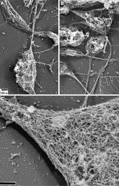
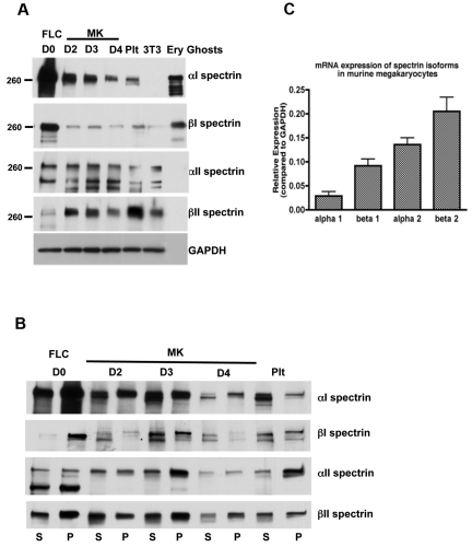
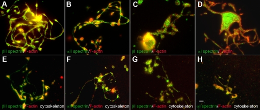
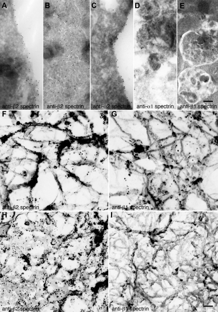
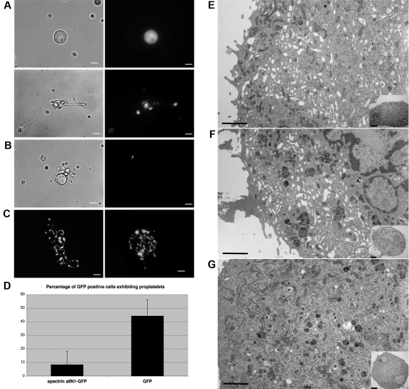
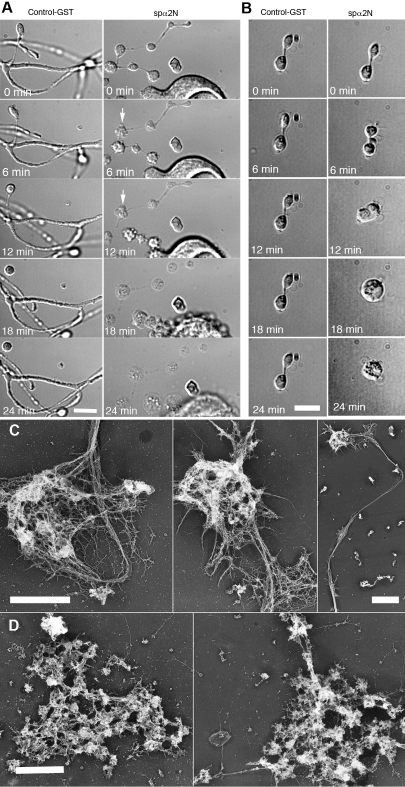
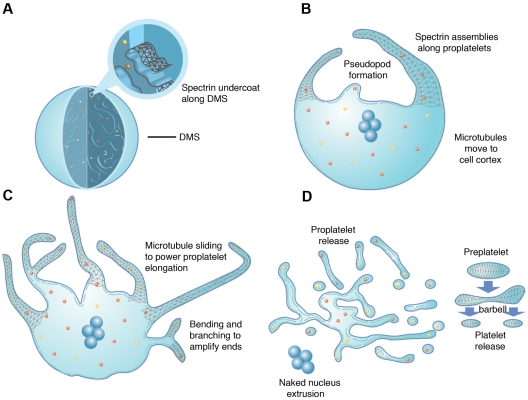
Megakaryocytes generate platelets by remodeling their cytoplasm first into proplatelets and then into preplatelets, which undergo fission to generate platelets. Although the functions of microtubules and actin during platelet biogenesis have been defined, the role of the spectrin cytoskeleton is unknown. We investigated the function of the spectrin-based membrane skeleton in proplatelet and platelet production in murine megakaryocytes. Electron microscopy revealed that, like circulating platelets, proplatelets have a dense membrane skeleton, the main fibrous component of which is spectrin. Unlike other cells, megakaryocytes and their progeny express both erythroid and nonerythroid spectrins. Assembly of spectrin into tetramers is required for invaginated membrane system maturation and proplatelet extension, because expression of a spectrin tetramer-disrupting construct in megakaryocytes inhibits both processes. Incorporation of this spectrin-disrupting fragment into a novel permeabilized proplatelet system rapidly destabilizes proplatelets, causing blebbing and swelling. Spectrin tetramers also stabilize the "barbell shapes" of the penultimate stage in platelet production, because addition of the tetramer-disrupting construct converts these barbell shapes to spheres, demonstrating that membrane skeletal continuity maintains the elongated, pre-fission shape. The results of this study provide evidence for a role for spectrin in different steps of megakaryocyte development through its participation in the formation of invaginated membranes and in the maintenance of proplatelet structure.


Figures

Figure 1
Figure 1
Structure of the proplatelet membrane skeleton. (A-B) Representative electron micrographs of the detergent-insoluble proplatelet cytoskeleton. Proplatelets were permeabilized with 0.75% Triton X-100, 0.1% glutaraldehyde, and 5μM phallacidin in PHEM buffer. Examination of the proplatelet membrane skeleton through electron microscopy reveals an intact membrane skeleton that laminates the underside and extends along the entire length of proplatelets. Inset: DIC image of murine proplatelets. Scale bar indicates 500 nm. (C) High-magnification, 3D electron micrograph of the proplatelet membrane skeleton showing the lattice-like network of elongated filamentous strands, which is similar in nature to the spectrin-based meshwork in erythrocytes and platelets. The membrane skeleton continuously laminates the underside of the proplatelet. A cytoplasmic bridge is shown (left) connecting to a swelling (right). Scale bar indicates 200 nm.
Figure 2
Figure 2
Spectrin isoforms in megakaryocytes and platelets. (A) Immunoblot showing the presence of erythroid and nonerythroid spectrin isoforms in MKs and platelet lysates. Isoform-specific antibodies were used to identify α1, β1, α2, and β2 spectrins in MKs at different stages of maturation and in platelets (Plt). Accordingly, α2 and β2 antibodies failed to recognize the erythroid spectrin isoforms in lysates of erythrocyte ghosts, whereas α1 and β1 antibodies identified the erythroid spectrin isoforms in the erythrocyte ghosts. Murine fibroblasts (3T3 Swiss) were used as a negative control for α1 and β1 spectrins. GAPDH was used as a loading control. Blots show anti-spectrin isoform labeling during different days of megakaryocyte culture: FLCs (day 0) and MKs at different stages: day 2, young MKs; day 3, MKs just before producing proplatelets; day 4, MKs after producing proplatelets. (B) Immunoblot analysis showing the distribution of spectrin isoforms in the pelleted (P) actin cytoskeleton and soluble (S) fractions of FLCs, MKs, and platelets. A higher fraction of αII and βII spectrin isoforms associated with the cytoskeletons in MKs just before making proplatelets (day 3), compared with other MK stages. Western blots from 3 different experiments were quantified by densitometry (supplemental Figure 6). (C) Quantitative PCR. The relative mRNA expression (compared with GAPDH) of spectrin isoforms in MKs determined by quantitative RT-PCR. Nonerythroid spectrins (α2 and β2 spectrins) were expressed at higher levels than erythroid isoforms (α1 and β1 spectrins) in MKs.
Figure 3
Figure 3
Localization of spectrin isoforms within proplatelets. Micrographs of immunofluorescence studies performed with spectrin antibodies reveal differential localizations for spectrin isoforms within proplatelets. The top panels (A-D) show proplatelet-producing MKs that were fixed before staining. The bottom panels (E-H) denoted “cytoskeleton,” show proplatelets permeabilized in 0.1% Triton-X 100 before fixation to remove soluble and membrane-associated structures. All micrographs are merged images of cells that were double-labeled with spectrin isoform antibodies (green) and F-actin staining by phalloidin (red). The isotype probed and the fluorophore of the secondary antibody are indicated in each panel. (A-D) α1 and β1 spectrins decorate punctate spots distributed throughout the proplatelets. α2 and β2 spectrins localized strongly along proplatelet shafts, proplatelet tips, and swellings. Both α2 and β2 spectrin isoforms colocalized with F-actin. All 4 spectrin isoforms tested were retained in the cytoskeleton of permeabilized cells (E-H) and displayed a similar localization pattern to nonextracted cells. Spectrin 1 isoforms stained in a punctate pattern throughout the proplatelet skeleton, whereas spectrin 2 isoforms displayed a more cytoskeletal localization in permeabilized cells. Scale bar indicates 5 μm.
Figure 4
Figure 4
Localization of spectrin isoforms at high resolution. (A-E) Localization of erythroid and nonerythroid spectrin isoforms in ultrathin sections of mouse MKs. Immunogold labeling of sections was performed with anti–β2 spectrin (A,B), anti–α2 spectrin (C), anti–α1 spectrin (D), and anti–β1 spectrin (E) antibodies. Gold particles (10-nm) recognizing anti–β2 spectrin (A-B) are evident on the plasma membrane and invaginated membranes of MKs. Gold particles recognizing anti–α2 spectrin (C) are also found on MK membranes. Gold particles recognizing anti–α1 spectrin (D) and anti–β1 spectrin (E) stained multivesicular bodies of MKs. Scale bar represents 200 nm. (F-I) Localization of spectrin isoforms in the detergent-insoluble cytoskeletons of proplatelets and platelets. Immunoelectron microscopic studies were used to localize individual spectrin isoforms in the membrane skeletons of proplatelets (F-G) and platelets (H-I). Preparations were incubated with affinity-purified anti-β2 (F,H) and anti–β1 spectrin (G,I) antibodies, followed by 10-nm gold particles coated with secondary antibodies. Scale bar shown in panel I indicates 100 nm. Proplatelets labeled with anti–β2 spectrin (F) and anti–β1 spectrin (G) have similar staining patterns, labeling the strands composing the membrane skeleton. Human platelet cytoskeletons were labeled with anti–β2 spectrin (H) and anti–β1 spectrin (I). β2 spectrin gold labeling was increased, whereas β1 was decreased in the platelet cytoskeleton. The gold particle density per square micrometer of cytoskeleton preparation was 53 ± 10 for anti–β2 spectrin of proplatelets (F), 69 ± 8 for anti–β1 spectrin of proplatelets (G) 232 ± 25 for anti–β2 spectrin of platelets (H), and 4 ± 2 for anti–β1 spectrin of platelets. Data are provided as means ± SE (n = 20). Experiments were carried out in triplicate. Scale bar represents 100 nm.
Figure 5
Figure 5
Expression of spα2N1 inhibits proplatelet elaboration by MKs and prevents IMS maturation. (A) The introduction of spα2N1-GFP in MKs through retroviral infection inhibited proplatelet formation. Most MKs expressing spα2N1-GFP failed to make proplatelets (A top panel), although a few developed primitive proplatelets (A bottom panel). Left panels show phase contrast images and right panels show fluorescence images. (B) Control uninfected MKs, identified by lack of green fluorescence, form normal proplatelets. (C) Fluorescence images of proplatelet formation by control MKs expressing GFP alone. Scale bars indicate 7.5 μm. (D) Quantitative analysis of proplatelet formation in spα2N1-GFP–expressing and control, GFP-expressing MKs. spα2N1-GFP–expressing MKs show a dramatic reduction in the percentage of proplatelets formed (8% compared with 44% in control cells). Bars represent the standard deviations. (E-G) Representative electron micrographs of a noninfected MK (E), a MK expressing GFP alone (F), and a MK expressing spα2N1-GFP (G). Control MKs (E-F) show an extensive, open IMS that fills the cell cytoplasm, whereas spα2N1-GFP–expressing MKs (G) do not. Insets show low-magnification views of the corresponding cells. Scale bars indicate 4μm.
Figure 6
Figure 6
Effect of spα2N1 on proplatelets. (A-B) Time-lapse DIC micrographs of permeabilized (0.4% OG) proplatelets treated with GST control polypeptide (left panels) and the spectrin-disrupting polypeptide spα2N1 (right panels) show that proplatelets treated with control GST maintain their “beads-on-a-string” structure and branches. In contrast, platelet-sized beads on proplatelets treated with spα2N1 blebbed and then underwent extensive swelling. After treatment with spα2N1, barbell-shaped proplatelets first blebbed and then fused their 2-platelet-sized swellings, forming a preplatelet-sized spheroid. (C-D) Electron micrographs of representative cytoskeletons from permeabilized proplatelets treated with either GST-control (C) or spα2N1 peptide (D). Scale bars indicate 5 μm. The cytoskeletons of GST-treated cells remain intact, whereas the cytoskeletons of spα2N1-treated cells are disrupted and aggregated.
Figure 7
Figure 7
Model of platelet production as suggested by present data and previous studies. As MKs transition from immature cells (A) to released platelets (D), a systematic series of events occurs. (A) MKs develop a highly IMS as they mature. Assembly of the spectrin-based membrane skeleton is involved in the formation of the IMS, providing a membrane reservoir for future formation of proplatelets. (B) Proplatelet production begins with the extension of large pseudopodia that use unique cortical bundles of microtubules to elongate and form thin proplatelet processes with bulbous ends. Proplatelet membranes are lined with a spectrin undercoat. Proplatelet termini contain a bundle of microtubules that loop on themselves. (C) Proplatelet elongation requires the sliding of microtubules past one another, driven by the molecular motor cytoplasmic dynein. As proplatelets elongate, expansion of the membrane surface area requires the outflow of the IMS, a process that likely requires remodeling of the membrane skeleton. Microtubules function as the highways on which mitochondria and granules traffic to the tips of proplatelets. Actin promotes the branching and amplification of proplatelet tips, representing a mechanism to increase the numbers of proplatelet ends, and ultimately, platelets. (D) The entire MK cytoplasm is converted into a mass of proplatelets and preplatelets (anucleate discoid particles 2-10 μm across), which are released from the cell. Preplatelets reversibly convert into barbell proplatelets, a process that is driven by twisting microtubule-based forces. The membrane skeleton stabilizes this barbell form. Platelets release from proplatelet ends after the final fission event. The nucleus is eventually extruded from the mass of proplatelets.

Comment in

Similar articles

Cited by

References

    1. Fox J, Boyles J, Berndt M, Steffen P, Anderson L. Identification of a membrane skeleton in platelets. J Cell Biol. 1988;106(5):1525–1538. - PMC - PubMed
    1. Fox J, Reynolds C, Morrow J, Phillips D. Spectrin is associated with membrane-bound actin filaments in platelets and is hydrolyzed by the Ca2+-dependent protease during platelet activation. Blood. 1987;69(2):537–545. - PubMed
    1. Hartwig J, DeSisto M. The cytoskeleton of the resting human blood platelet: Structure of the membrane skeleton and its attachment to actin filaments. J Cell Biol. 1991;112(3):407–425. - PMC - PubMed
    1. Byers TJ, Branton D. Visualization of the protein associations in the erythrocyte membrane skeleton. Proc Nat Acad Sci U S A. 1985;82(18):6153–6157. - PMC - PubMed
    1. Fox J, Aggerbeck L, Berndt M. Structure of the glycoprotein Ib-IX complex from platelet membranes. J Biol Chem. 1988;263(10):4882–4890. - PubMed

Publication types

MeSH terms