. 2011 Jul 8;286(27):23742-52.
doi: 10.1074/jbc.M111.220780. Epub 2011 May 12.

Molecular insight into how HIV-1 Vpr protein impairs cell growth through two genetically distinct pathways

Affiliations

Molecular insight into how HIV-1 Vpr protein impairs cell growth through two genetically distinct pathways

Claire Maudet et al. J Biol Chem. .

Abstract

Vpr, a small HIV auxiliary protein, hijacks the CUL4 ubiquitin ligase through DCAF1 to inactivate an unknown cellular target, leading to cell cycle arrest at the G(2) phase and cell death. Here we first sought to delineate the Vpr determinants involved in the binding to DCAF1 and to the target. On the one hand, the three α-helices of Vpr are necessary and sufficient for binding to DCAF1; on the other hand, nonlinear determinants in Vpr are required for binding to the target, as shown by using protein chimeras. We also underscore that a SRIG motif conserved in the C-terminal tail of Vpr proteins from HIV-1/SIVcpz and HIV-2/SIVsmm lineages is critical for G(2) arrest. Our results suggest that this motif may be predictive of the ability of Vpr proteins from other SIV lineages to mediate G(2) arrest. We took advantage of the characterization of a subset of G(2) arrest-defective, but DCAF1 binding-proficient mutants, to investigate whether Vpr interferes with cell viability independently of its ability to induce G(2) arrest. These mutants inhibited cell colony formation in HeLa cells and are cytotoxic in lymphocytes, unmasking a G(2) arrest-independent cytopathic effect of Vpr. Furthermore these mutants do not block cell cycle progression at the G(1) or S phases but trigger apoptosis through caspase 3. Disruption of DCAF1 binding restored efficiency of colony formation. However, DCAF1 binding per se is not sufficient to confer cytopathicity. These data support a model in which Vpr recruits DCAF1 to induce the degradation of two host proteins independently required for proper cell growth.


Figures

FIGURE 1.
FIGURE 1.
The C-terminal tail of Vpr shows a highly conserved SRIG motif, which is critical for cell cycle arrest activity. A, identification of a highly conserved SRIG motif in the C-terminal tail of Vpr from HIV-1/SIVcpz and HIV-2/SIVsmm/SIVmnd2/SIVrcm is shown. The Vpr and Vpx proteins with the indicated accession numbers were aligned from the end of the predicted third α-helix as defined by the underlined invariant GC di-amino acid motif to their C termini. The lentiviral origin of the proteins is indicated on the left side. The conserved SRIG motif is boxed. B, the Vpr proteins from SIVmnd-2 and SIVrcm both bind DCAF1. Cell lysates were prepared 48 h post-transfection and subjected to immunoprecipitation using anti-FLAG antibodies covalently coupled to Sepharose beads. After extensive washing, bound proteins were eluted from beads with the FLAG peptide. Immunoprecipitates (IP) and crude cell lysates (Lysates) were analyzed by Western blotting using the indicated antibodies. C, SIVmnd-2 Vpr and SIVrcm Vpr arrest the cell cycle similarly to HIV-1 Vpr. HeLa cells were transfected with vectors expressing the indicated HA-tagged proteins along with a vector expressing the GFP protein. Cells were harvested 48 h post-transfection. After fixation and propidium iodide staining, the cells were analyzed by flow cytometry to monitor the DNA content of the GFP-positive population. The G2/G1 ratio is indicated in the bar graph. D, each residue of the SRIG motif is critical for the cell cycle arrest activity of HIV-1 Vpr. Alanine substitutions of each residue of the SRIG motif were introduced into the Vpr protein from HIV-1 LAI by site-directed mutagenesis. The Vpr-mutated proteins were expressed into HeLa cells to monitor their effect on cell cycle progression in comparison to wt Vpr. Cell cycle analysis was performed as described above (top). A fraction of cells was lysed, and the indicated HA-tagged Vpr proteins as well as the co-transfected GFP protein were revealed by Western blot analysis. Quantification of the signals enabled to deduce the Vpr to GFP protein expression ratio that is represented on the bar graph (bottom).
FIGURE 2.
FIGURE 2.
Chimeric proteins between a DCAF1 binding module and the C-terminal tail of HIV-1 Vpr are defective for G2 arrest activity. A, the three-α-helices core domain of HIV-1 Vpr and SIVsmm Vpx is necessary and sufficient to bind DCAF1. The indicated hybrid proteins were co-expressed in the L40 yeast reporter strain, and the yeast transformants were analyzed for induction of β-galactosidase (left panel). As a control, the right panel shows the normal growth in permissive medium of the corresponding yeast transformants. B, the structure of the HIV-1 Vpr, SIVsmm Vpx, and SIVagm Vpr proteins and of their chimeric X-R and Ragm-R derivatives is shown. The viral proteins are depicted as rectangles. Their predicted three α-helices, hereafter denoted α1, α2, and α3, are represented by dotted and hatched boxes. Shown are the positions of several residues fully conserved between the three proteins including the Tyr and His residues in α1, the Gln and His residues in α3, and the GC di-amino acid at the boundary between α3 and the C-terminal tail. The Gln residue, which is critical for DCAF1 binding, is underlined. The SRIG motif found in the C-terminal tail of HIV-1 Vpr as well as the PPGLD motif found in the C-terminal tail of SIVagm Vpr and a conserved G-P rich motif found in the C-terminal tail of SIVsmm Vpx are indicated. C, X-R and Ragm-R chimera both bind DCAF1. Co-immunoprecipitation (IP) experiments were performed as described in Fig. 1B. D, fusion of the C-terminal tail of HIV-1 Vpr to the DCAF1-binding region of SIVsmm Vpx or SIVagm Vpr fails to restore the G2 arrest activity of wt Vpr. HeLa cells expressing the indicated constructs were analyzed for their cell cycle profile as described in Fig. 1C.
FIGURE 3.
FIGURE 3.
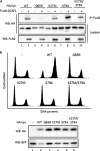
Vpr mutations lying outside of the C-terminal tail abrogate G2 arrest activity while preserving DCAF1 binding function. A, the K27M and K27M/S79A Vpr mutants efficiently co-immunoprecipitate (IP) with DCAF1. Constructs expressing the indicated proteins were transfected into HeLa cells. Cell lysates were prepared 48 h post-transfection and subjected to immunoprecipitation as described in Fig. 1B. Note that the VprQ65R mutant is unable to co-immunoprecipitate with DCAF1 consistent with our previously published results (23). WB, immunoblot. B, the K27M Vpr mutant fails to promote G2 arrest. HeLa cells expressing the indicated proteins were analyzed for their cell cycle profiles and for protein expression as described in Fig. 1C.
FIGURE 4.
FIGURE 4.
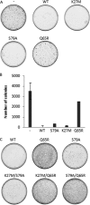
G2 arrest-defective mutants of Vpr conserve cytotoxicity in a DCAF1 binding-dependent manner. HeLa cells were transfected in duplicate wells of 12-well plates with EBV-based episomes expressing the indicated proteins. Two days after transfection, cells were harvested, and 1:10 of the cell population was transferred into 10-cm dishes and grown in hygromycin-containing medium. The formation of hygromycin-resistant colonies due to stable maintenance of the episome was visualized by Giemsa staining 15 days post-selection. The result of a representative experiment is shown in A, and quantification of six independent experiments is illustrated in the bar graph in B. The results of an additional experiment including three new double mutants are presented in C.
FIGURE 5.
FIGURE 5.
The DCAF1 binding activity of the Vpr/Vpx superfamily is not sufficient to confer cytopathicity. A, Vpx from SIVsmm and Vpr from SIVagm bind DCAF1 as efficiently as HIV-1 Vpr. The indicated constructs were transfected into HeLa cells. Cell lysates were prepared 48 h post-transfection and subjected to immunoprecipitation (IP) as described in Fig. 1B. WB, immunoblot. B, neither Vpx from SIVsmm nor Vpr from SIVagm can mediate cytopathicity. The colony formation assay was performed as described for Fig. 4.
FIGURE 6.
FIGURE 6.
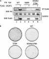
UNG2 does not mediate Vpr-induced cytopathicity in the clonogenic assay. A, the UNG2 binding-deficient W54R/S79A Vpr mutant is still able to recruit DCAF1. The indicated constructs were transfected into HeLa cells. Immunoprecipitation (IP) was performed as described for Fig. 1B. WB, immunoblot. B, preventing binding between Vpr and UNG2 does not alter the cytopathic activity of Vpr. The colony formation assay was performed as described for Fig. 4.
FIGURE 7.
FIGURE 7.
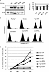
G2 arrest-independent cytotoxicity of Vpr correlates with induction of apoptosis but not with a block in cell cycle progression. A, the expression of VprK27M or VprS79A induces an accumulation of cells in the sub-G1 phase. HeLa cells were transfected with the indicated EBV constructs. 3, 4, and 5 days post-selection in hygromycin-containing medium, cells were harvested and analyzed for their cell cycle profiles as described in Fig. 1 legend. Flow cytometry analysis was performed with the total cell population to include cells with a DNA content <2N (sub-G1), indicative of an ongoing apoptotic death. The bar graph represents the proportion of cells in sub-G1 over time. B, expression of VprK27M and VprS79A mediate activation of caspase 3 is shown. HeLa cells were transfected with the indicated EBV constructs. 4 days post-selection in hygromycin-containing medium, cells were harvested and lysed, and proteolytic activation of caspase 3 was analyzed by a Western blot. C, the long term cytopathic activity of Vpr is independent of a cell cycle blockage. HeLa cells were transfected with the indicated EBV constructs. After 3 days in hygromycin-containing medium, cells were incubated for 1 h with EdU, a BrdU analog that is incorporated into DNA during the S phase. Cells were then fixed, stained with propidium iodide, and analyzed by flow cytometry. The cell cycle status of the cells as presented at the top is explained by the small graph in the middle. The bar graph at the bottom represents for each Vpr mutant the percentage of cells in the different phases of the cell cycle.
FIGURE 8.
FIGURE 8.
Vpr from the incoming virion induces apoptosis independently of its G2 arrest activity in Jurkat cells. A, Vpr wt and mutants are similarly incorporated into HIV-1 pseudoparticles. Pseudoparticles were produced in the absence or presence of HA-tagged wt or mutant Vpr proteins. The presence of Vpr and the p24 capsid in the viral particle was analyzed by Western blot (left panel). The signals obtained in the left panel were quantified and expressed as HA/p24 ratios (right panel). B and C, VprK27M and VprS79A, but not VprQ65R, induce apoptosis in Jurkat cells. Jurkat cells were incubated with empty VLP or VLP containing the indicated HA-Vpr proteins each day until harvesting. Cells were harvested 48, 72, and 96 h after the first incubation, and the number of apoptotic cells was determined by flow cytometry after labeling with annexin-FITC. Panel B shows the distribution of cells versus the staining intensity at 96h. Panel C shows the percentage of annexin-FITC-positive cells versus time.

References

    1. Malim M. H., Emerman M. (2008) Cell Host Microbe 3, 388–398 - PubMed
    1. Chakrabarti L., Guyader M., Alizon M., Daniel M. D., Desrosiers R. C., Tiollais P., Sonigo P. (1987) Nature 328, 543–547 - PubMed
    1. Lang S. M., Weeger M., Stahl-Hennig C., Coulibaly C., Hunsmann G., Müller J., Müller-Hermelink H., Fuchs D., Wachter H., Daniel M. M. (1993) J. Virol. 67, 902–912 - PMC - PubMed
    1. Balliet J. W., Kolson D. L., Eiger G., Kim F. M., McGann K. A., Srinivasan A., Collman R. (1994) Virology 200, 623–631 - PubMed
    1. Connor R. I., Chen B. K., Choe S., Landau N. R. (1995) Virology 206, 935–944 - PubMed

Publication types

Substances