. 2011 Jul 1;286(26):23570-81.
doi: 10.1074/jbc.M111.230235. Epub 2011 May 12.

Transport mechanism and pH regulation of the Na+/H+ antiporter NhaA from Escherichia coli: an electrophysiological study

Affiliations

Transport mechanism and pH regulation of the Na+/H+ antiporter NhaA from Escherichia coli: an electrophysiological study

Thomas Mager et al. J Biol Chem. .

Abstract

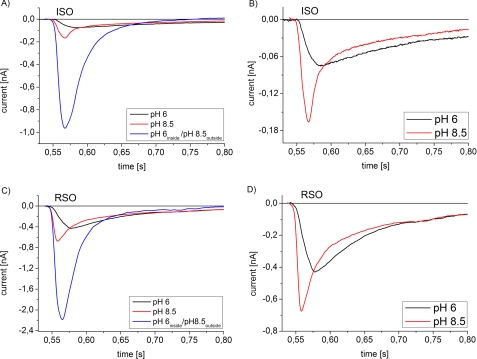
Using an electrophysiological assay the activity of NhaA was tested in a wide pH range from pH 5.0 to 9.5. Forward and reverse transport directions were investigated at zero membrane potential using preparations with inside-out and right side-out-oriented transporters with Na(+) or H(+) gradients as the driving force. Under symmetrical pH conditions with a Na(+) gradient for activation, both the wt and the pH-shifted G338S variant exhibit highly symmetrical transport activity with bell-shaped pH dependences, but the optimal pH was shifted 1.8 pH units to the acidic range in the variant. In both strains the pH dependence was associated with a systematic increase of the K(m) for Na(+) at acidic pH. Under symmetrical Na(+) concentration with a pH gradient for NhaA activation, an unexpected novel characteristic of the antiporter was revealed; rather than being down-regulated, it remained active even at pH as low as 5. These data allowed a transport mechanism to advance based on competing Na(+) and H(+) binding to a common transport site and a kinetic model to develop quantitatively explaining the experimental results. In support of these results, both alkaline pH and Na(+) induced the conformational change of NhaA associated with NhaA cation translocation as demonstrated here by trypsin digestion. Furthermore, Na(+) translocation was found to be associated with the displacement of a negative charge. In conclusion, the electrophysiological assay allows the revelation of the mechanism of NhaA antiport and sheds new light on the concept of NhaA pH regulation.


Figures

FIGURE 1.
FIGURE 1.
Transport modes of NhaA in the bacterial cell and under experimental conditions. The left column shows the forward transport mode of NhaA corresponding to its physiological transport direction in E. coli, and the right column shows the reverse transport mode. In the upper panel a graphic representation of both transport modes in the bacterial cell is given. The lower two panels show the experimental preparations used for the investigation of the two transport modes. Na+ gradient (ΔNa)- or pH gradient (ΔpH)-driven transport activity of NhaA and the corresponding transient currents observed on the SSM are schematically depicted. Note the different polarity of the currents at different conditions.
FIGURE 2.
FIGURE 2.
pH dependence of transient currents obtained with wt- and G338S-NhaA after a Na+ concentration jump using the single solution exchange protocol. A and B, typical transient currents were recorded after a 100 mm Na+ concentration jump using RSO proteoliposomes and ISO membrane vesicles. Signals of ISO membrane vesicles are a superposition of the NhaA transporter current (negative phase) and the solution exchange artifact (positive phase). Signals of RSO proteoliposomes are so large that the contribution of artifacts is negligible. C and D, normalized peak currents at the indicated pH values were subtracted by the Na+ jump artifact. E and F, shown are normalized reconstructed stationary currents at the indicated pH values. The graphs show average values from recordings using three individual sensors and the corresponding S.D. Currents are normalized as described under “Experimental Procedures.” A, C, and E, ISO membrane vesicles with G338S NhaA (red) or wt NhaA (black) are shown. B, D, and F, RSO proteoliposomes (LPR = 10) with G338S NhaA (red) or wt NhaA (black) are shown. For the Na+ concentration jumps, activating solutions containing 10 mm NaCl and 290 mm KCl or 100 mm NaCl and 200 mm KCl titrated to the indicated pH values with HCl or Tris were used. The nonactivating solutions contained 300 mm KCl instead. In addition all buffers contained 5 mm MgCl2, 25 mm Tris, 25 mm MOPS, 25 mm MES, and 1 mm DTT.
FIGURE 3.
FIGURE 3.
Na+ concentration dependence of wt NhaA at optimal and acidic pH. The transient currents were recorded after a Na+ concentration jump using the single solution exchange protocol. A and B, normalized peak currents with ISO membrane vesicles are shown. C and D, normalized peak currents with RSO proteoliposomes (LPR = 10) are shown. The graphs show average values from recordings using three individual sensors and the corresponding S.D. Normalization was as described under “Experimental Procedures.” For the Na+ concentration jumps, activating solutions containing x mm NaCl and (300–x) mm KCl titrated to the indicated pH values with Tris were used. The nonactivating solutions contained 300 mm KCl instead. In addition, all buffers contained 5 mm MgCl2, 25 mm Tris, 25 mm MOPS, 25 mm MES, and 1 mm DTT. The solid line is a fit to the data using a hyperbolic model function with a half-saturation concentration Km and a fixed vmax = 1.
FIGURE 4.
FIGURE 4.
Na+ concentration dependence of G338S NhaA at optimal and acidic pH. The transient currents were recorded after a Na+ concentration jump using the single solution exchange protocol. A and B, normalized peak currents with ISO membrane vesicles are shown. C and D, normalized peak currents with RSO proteoliposomes (LPR = 10) are shown. The graphs show average values from recordings using three individual sensors and the corresponding S.D. Normalization was as described under “Experimental Procedures.” For the Na+ concentration jumps, activating solutions containing x mm NaCl and (300–x) mm KCl titrated to the indicated pH values with Tris were used. The nonactivating solutions contained 300 mm KCl instead. In addition, all buffers contained 5 mm MgCl2, 25 mm Tris, 25 mm MOPS, 25 mm MES, and 1 mm DTT. The solid line is a fit to the data using a hyperbolic model function with a half-saturation concentration Km and a fixed vmax = 1.
FIGURE 5.
FIGURE 5.
Transient currents after a pH jump using wt NhaA RSO proteoliposomes (LPR = 50). The currents were measured by means of the single solution exchange protocol. A, for comparison, a ΔNa (100 mm) signal was recorded using the same sensor (upper trace). The ΔpH signals (lower traces) show a solution exchange from pH 8.5 to 5.0 or from pH 5.0 to 8.5 at a symmetrical Na+ concentration of 100 mm. The circle represents the liposome, and the numbers represent the pH inside and outside. For comparison, the corresponding control signal with K+ instead of Na+ is given (dashed lines). B, shown are peak currents at different pH values and a symmetrical Na+ concentration of 100 mm normalized to the ΔNa peak current shown in A. Solution exchange from pH 8.5 to the indicated pH is shown. C, shown are peak currents at different pH values and a symmetrical Na+ concentration of 100 mm normalized to the ΔNa peak current shown in A. Solution exchange from the indicated pH to pH 8.5 is shown. D and E, KmNA for the measurements shown in panels (F, G, H, I, J, and K). Panels F, G, H, I, J, and K show the normalized peak currents after a pH jump at different symmetrical Na+ concentrations. Average values from recordings using 3 individual sensors and the corresponding S.D. are displayed. Normalization is as described under “Experimental Procedures.” For measurement conditions, the ΔNa signal was recorded using the conditions described in Fig. 2 for 100 mm Na+ and pH 8.5. For the ΔpH signals, the solutions contained x mm NaCl and (300–x) mm KCl. In addition, buffers (F–I) contained 5 mm MgCl2, 25 mm Tris, 25 mm MOPS, 25 mm MES, and 1 mm DTT. Buffers (J and K) additionally contained 5 mm MgCl2, 25 mm Tris, 25 mm MOPS, 25 mm citrate, and 1 mm DTT.
FIGURE 6.
FIGURE 6.
Transient currents of G338S NhaA at alkaline pH. For comparison, the transient currents after a 10 mm Na+ concentration jump at a symmetric pH of 6 (black) and 8.5 (red) were measured with a single solution exchange protocol. For the pH gradient measurements (blue), the double solution exchange protocol was used. A, G338S ISO membrane vesicles are shown. C, G338S RSO proteoliposomes (LPR = 10) are shown. For the Na+ concentration jumps, activating solutions containing 10 mm NaCl and 290 mm KCl titrated to pH 6 (black) or pH 8.5 (red and blue) with HCl or Tris were used. The nonactivating solutions contained 300 mm KCl titrated to the same pH as the corresponding activating solutions. The resting solutions for the establishment of the ΔpH (blue) contained 300 mm KCl at pH 6 (blue). In addition, all buffers contained 5 mm MgCl2, 25 mm Tris, 25 mm MOPS, 25 mm MES, and 1 mm DTT. B and D, shown are the same data as in A and C. For a better comparison, an expanded view of the transient currents is given.
FIGURE 7.
FIGURE 7.
Na+ and pH dependence of tryptic digestion of wt NhaA. Trypsin digestion of purified NhaA was conducted in the absence (lanes 1, 4, and 7) or presence of 10 mm Na+ (lanes 2, 5, and 8) and 100 mm Na+ (lanes 3, 6, and 9) at the indicated pH values. A, the digestion products were resolved on SDS-PAGE with a control of undigested NhaA (lane 10). B, the extent of digestion was determined by analysis of the density of the gel bands shown in A. The experiment was repeated three times with practically identical results.
FIGURE 8.
FIGURE 8.
Minimal kinetic model used for the analysis of the transport properties of NhaA. Outward-facing transporter Co binds substrates either H+o or Na+o from the outward (periplasmic) medium, and inward facing carrier Ci binds substrates either H+i or Na+i from the inward (cytoplasmic) medium. The schematics show the proposed transport mechanism (see “Discussion”). The conformational transitions CoH⇋CiH and CoNa⇋CiNa are associated with a displacement of the two negatively charged aspartate residues (Asp-163, Asp-164) and one Na+ or two H+ ions, respectively. In the case of Na+ translocation, this leads to a net charge displacement as indicated by red arrows. Note that in the crystal structure obtained at pH 4.0 Asp-163 is occluded.
FIGURE 9.
FIGURE 9.
Simultaneous fit of the pH dependence and the sodium dependence of the peak currents to the minimal kinetic model. The graph shows sodium jump-induced peak currents of wt NhaA and G338S NhaA in native transport direction (A, B, E, and F) and reverse transport direction (C, D, G, and H). Data and conditions are as in Figs. 2–4. The solid line is a fit to the minimal kinetic model described in the “Discussion.” The kinetic parameters obtained by the fit are given in the figure.

Similar articles

Cited by

References

    1. Padan E., Bibi E., Ito M., Krulwich T. A. (2005) Biochim. Biophys. Acta 1717, 67–88 - PMC - PubMed
    1. Brett C. L., Donowitz M., Rao R. (2005) Am. J. Physiol. Cell Physiol. 288, 223C–239C - PubMed
    1. Taglicht D., Padan E., Schuldiner S. (1991) J. Biol. Chem. 266, 11289–11294 - PubMed
    1. Padan E., Kozachkov L., Herz K., Rimon A. (2009) J. Exp. Biol. 212, 1593–1603 - PubMed
    1. Rothman A., Gerchman Y., Padan E., Schuldiner S. (1997) Biochemistry 36, 14572–14576 - PubMed

Publication types

MeSH terms

LinkOut - more resources