. 2011 Jul 8;286(27):23888-99.
doi: 10.1074/jbc.M111.245969. Epub 2011 May 12.

Molecular basis of secretin docking to its intact receptor using multiple photolabile probes distributed throughout the pharmacophore

Affiliations

Molecular basis of secretin docking to its intact receptor using multiple photolabile probes distributed throughout the pharmacophore

Maoqing Dong et al. J Biol Chem. .

Abstract

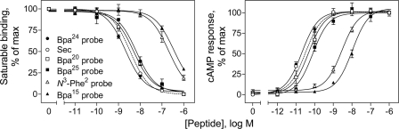
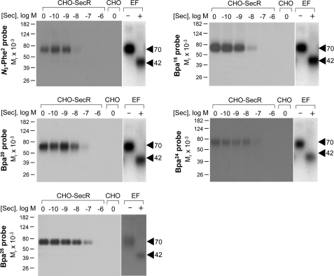
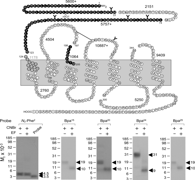
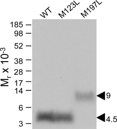
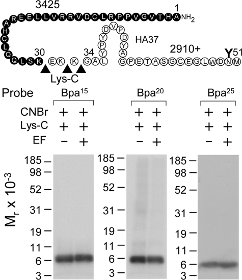
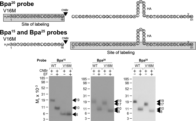
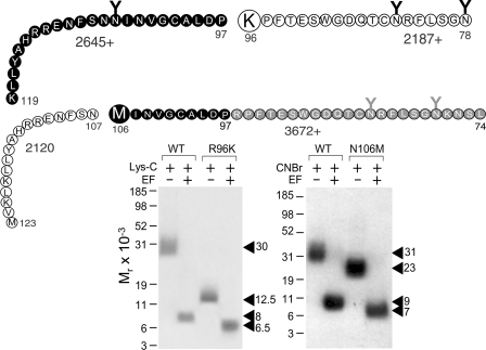
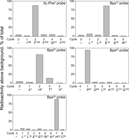
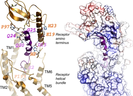
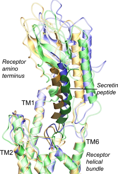
The molecular basis of ligand binding and activation of family B G protein-coupled receptors is not yet clear due to the lack of insight into the structure of intact receptors. Although NMR and crystal structures of amino-terminal domains of several family members support consistency in general structural motifs that include a peptide-binding cleft, there are variations in the details of docking of the carboxyl terminus of peptide ligands within this cleft, and there is no information about siting of the amino terminus of these peptides. There are also no empirical data to orient the receptor amino terminus relative to the core helical bundle domain. Here, we prepared a series of five new probes, incorporating photolabile moieties into positions 2, 15, 20, 24, and 25 of full agonist secretin analogues. Each bound specifically to the receptor and covalently labeled single distinct receptor residues. Peptide mapping of labeled wild-type and mutant receptors identified that the position 15, 20, and 25 probes labeled residues within the distal amino terminus of the receptor, whereas the position 24 probe labeled the amino terminus adjacent to TM1. Of note, the position 2 probe labeled a residue within the first extracellular loop of the receptor, a region not previously labeled, providing an important new constraint for docking the amino-terminal region of secretin to its receptor core. These additional experimentally derived constraints help to refine our understanding of the structure of the secretin-intact receptor complex and provide new insights into understanding the molecular mechanism for activation of family B G protein-coupled receptors.


Figures

FIGURE 1.
FIGURE 1.
Functional characterization of photolabile secretin analogues. Left, binding curves of increasing concentrations of secretin and each of the photolabile secretin probes to compete for binding of the secretin-like radioligand [125I-Tyr10]rat secretin-27 to secretin receptor-bearing CHO-SecR cells. The values illustrated represent saturable binding as percentages of maximal binding observed in the absence of the competing secretin and are expressed as the means ± S.E. of duplicate values from a minimum of three independent experiments. Right, curves of intracellular cAMP responses in CHO-SecR cells stimulated by increasing concentrations of secretin and each of the noted probes. Data points represent the means ± S.E. of three independent experiments performed in duplicate, normalized relative to the maximal response to secretin. Basal (5.7 ± 1.3 pmol/million cells) and maximal (189 ± 38 pmol/million cells) cAMP levels by each of the peptides were similar. Sec, secretin.
FIGURE 2.
FIGURE 2.
Photoaffinity labeling of secretin receptor. Shown are representative autoradiographs of 10% SDS-polyacrylamide gels used to separate the products of affinity labeling membranes from CHO-SecR cells with each of the noted photolabile probes in the presence of increasing concentrations of competing unlabeled secretin (Sec; from 0 to 1 μm). As controls, labeling of the non-receptor-bearing CHO cell membranes by each probe in the absence of competitor is also shown. Each of the probes labeled the secretin receptor specifically and saturably with the labeling being competed by secretin in a concentration-dependent manner. The receptor bands labeled by each probe migrated at approximately Mr = 70,000 and shifted to approximately Mr = 42,000 after deglycosylation with endoglycosidase F (EF). No radioactive band was observed in the affinity-labeled non-receptor-bearing CHO cell membranes. Data are representative of at least three independent experiments.
FIGURE 3.
FIGURE 3.
CNBr cleavage of labeled secretin receptor. Top, diagram illustrating the theoretical fragments of the secretin receptor resulting from CNBr cleavage. Bottom, results of CNBr cleavage of the secretin receptor labeled by each of the noted probes. Cleavage of the secretin receptor labeled by the position 2 probe yielded a non-glycosylated fragment migrating at approximately Mr = 4,500, representing either the fragment Tyr124–Met134 (beginning of TM1; gray circles) or Ile198–Met205 (beginning of TM3; black circles). Cleavage of the secretin receptor labeled by the position 15, 20, and 25 probes each yielded a fragment migrating at approximately Mr = 19,000 that shifted to approximately Mr = 10,000 after deglycosylation by endoglycosidase F (EF), likely representing the first CNBr fragment between Ala1 and Met51 (20). Cleavage of the secretin receptor labeled by the position 24 probe yielded a fragment migrating at approximately Mr = 31,000 that shifted to approximately Mr = 9,000 after deglycosylation, likely representing the third CNBr fragment between Leu74 and Met123 (21, 43). Data are representative of at least three independent experiments.
FIGURE 4.
FIGURE 4.
CNBr cleavage of mutant secretin receptors labeled by position 2 probe. CNBr cleavage of the labeled M197L mutant receptor resulted in a radioactive band migrating at approximately Mr = 9,000, clearly distinct from the migration pattern of the CNBr cleavage products from the wild-type (WT) and M123L mutant receptors (Mr = 4,500). This indicates that the Ile198–Met205 fragment spanning ECL1 and TM3 contained the site of labeling by the position 2 probe (see diagram in Fig. 3).
FIGURE 5.
FIGURE 5.
Lys-C cleavage of secretin receptor labeled by each of position 15, 20, and 25 probes. Top, diagram of the sites of Lys-C cleavage of the first CNBr fragment (Ala1–Met51) of the secretin receptor along with the masses of the protein cores of the resultant fragments. Bottom, Lys-C cleavage of the CNBr fragment of the secretin receptor labeled by each probe resulted in a non-glycosylated band migrating at approximately Mr = 6,000, representing the labeling of the Ala1–Lys30 fragment at the distal amino terminus of the receptor. EF, endoglycosidase F.
FIGURE 6.
FIGURE 6.
CNBr cleavage of V16M secretin receptor labeled by position 15, 20, and 25 probes. Top, diagrams of the sites of CNBr cleavage of the Ala1–Met51 region of the V16M mutant secretin receptor labeled by each probe. Bottom, CNBr cleavage of the V16M mutant receptor labeled by the position 20 probe yielded a non-glycosylated band migrating at approximately Mr = 6,000, representing the labeling of the amino-terminal Ala1–Met16 fragment. CNBr cleavage of the V16M mutant receptor labeled by position 15 and 25 probes each yielded a fragment migrating at approximately Mr = 17,000 that shifted to approximately Mr = 8,000 after deglycosylation, representing the labeling of the carboxyl-terminal Leu17–Met51 fragment. EF, endoglycosidase F.
FIGURE 7.
FIGURE 7.
Cleavage of R96K and N106M secretin receptor mutants labeled by position 24 probe. Top, diagrams of the sites of Lys-C and CNBr cleavage of the R96K and N106M mutant secretin receptor constructs, respectively, along with the masses of the protein cores of the resultant fragments. Bottom, Lys-C cleavage of the wild-type secretin receptor (WT) and the R96K mutant labeled by the position 24 probe resulted in bands migrating at approximately Mr = 30,000 and Mr = 12,500 that further shifted to approximately Mr = 8,000 and Mr = 6,500 after deglycosylation, respectively, indicating that the site of labeling was within the Pro97–Met123 fragment (21, 43). CNBr cleavage of the labeled N106M receptor yielded a band migrating at approximately Mr = 23,000 and shifting to approximately Mr = 7,000, distinct from the pattern of cleavage of the wild-type secretin receptor (approximate Mr = 31,000 shifting to approximate Mr = 9,000 after deglycosylation), indicating that the fragment Leu74–Asn106 contained the site of labeling (21). Taken together, the site of labeling with the position 24 probe was within the region between Pro97 and Asn106 of the secretin receptor. EF, endoglycosidase F.
FIGURE 8.
FIGURE 8.
Identification of labeled receptor residues by radiochemical sequencing. Shown are radioactive elution profiles of Edman degradation sequencing of the purified receptor fragments labeled by each of the position 2 (Ile198–Met205), 15 (Leu17–Met51), 20 (Ala1–Met51), 24 (Pro97–Lys119), and 25 (Leu17–Met51) probes. There were peaks in eluted radioactivity in cycles 2, 3, 2, 1, and 7 that correspond with covalent attachment to receptor residues Leu199, Glu19, His2, Pro97, and His23 by position 2, 15, 20, 24, and 25 probes, respectively.
FIGURE 9.
FIGURE 9.
Molecular model of ligand-bound secretin receptor. Shown is a lateral view of the best model of the secretin receptor-secretin peptide complex with the amino-terminal domain of the receptor above and the transmembrane helical bundle domain below. The secretin peptide ligand is illustrated in magenta with its carboxyl terminus within the peptide-binding cleft at the top and its amino terminus extending into the helical bundle at the bottom. The left panel highlights the five pairs of residues contributing to the experimental spatial approximation constraints (residues linked with blue dotted lines). The sites of incorporation of Bpa into the peptide ligand are identified in magenta, and the sites of covalent labeling of the receptor are identified in orange. The right panel illustrates the surfaces of the peptide-binding cleft within the receptor amino terminus and extending to the helical bundle.
FIGURE 10.
FIGURE 10.
Three best molecular models of ligand-bound secretin receptor. Shown is the high degree of structural similarity among the three best molecular models, superimposed by the backbone of their transmembrane domains. Model 1 is colored gold, model 2 is green, and model 3 is blue (in each model, the receptor is in lighter shade, whereas the peptide is darker).

References

    1. Ulrich C. D., 2nd, Holtmann M., Miller L. J. (1998) Gastroenterology 114, 382–397 - PubMed
    1. Cherezov V., Rosenbaum D. M., Hanson M. A., Rasmussen S. G., Thian F. S., Kobilka T. S., Choi H. J., Kuhn P., Weis W. I., Kobilka B. K., Stevens R. C. (2007) Science 318, 1258–1265 - PMC - PubMed
    1. Jaakola V. P., Griffith M. T., Hanson M. A., Cherezov V., Chien E. Y., Lane J. R., Ijzerman A. P., Stevens R. C. (2008) Science 322, 1211–1217 - PMC - PubMed
    1. Scheerer P., Park J. H., Hildebrand P. W., Kim Y. J., Krauss N., Choe H. W., Hofmann K. P., Ernst O. P. (2008) Nature 455, 497–502 - PubMed
    1. Warne T., Serrano-Vega M. J., Baker J. G., Moukhametzianov R., Edwards P. C., Henderson R., Leslie A. G., Tate C. G., Schertler G. F. (2008) Nature 454, 486–491 - PMC - PubMed

Publication types

MeSH terms

LinkOut - more resources