. 2011 May 13;332(6031):816-21.
doi: 10.1126/science.1202617.

Computational design of proteins targeting the conserved stem region of influenza hemagglutinin

Affiliations

Computational design of proteins targeting the conserved stem region of influenza hemagglutinin

Sarel J Fleishman et al. Science. .

Abstract

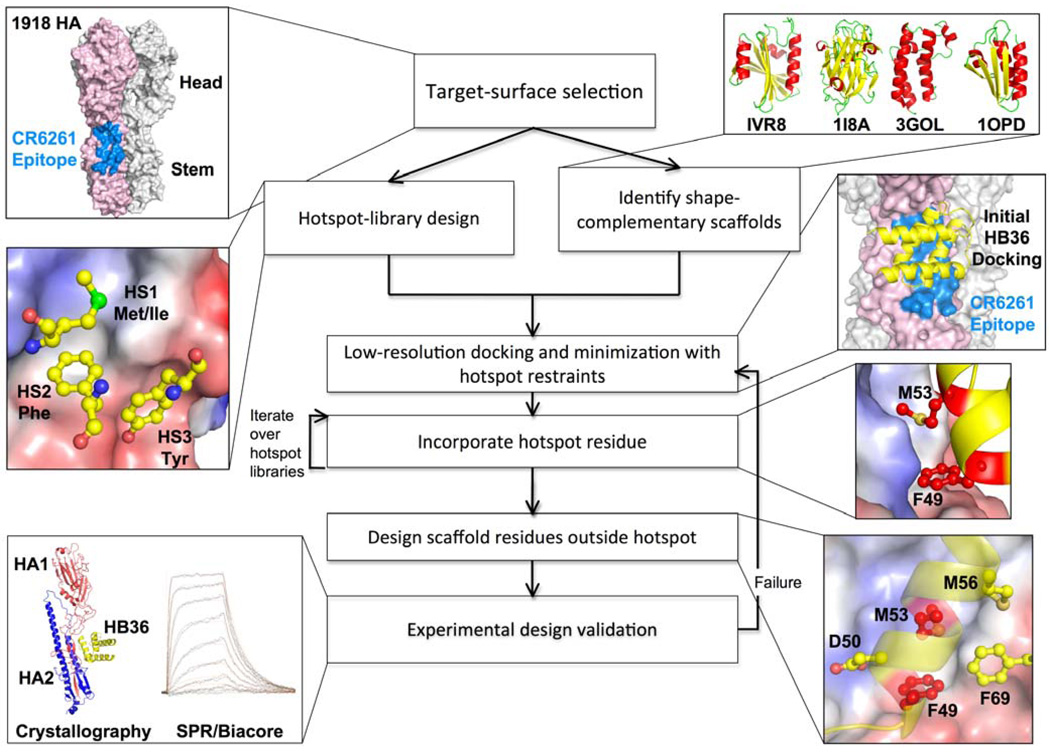
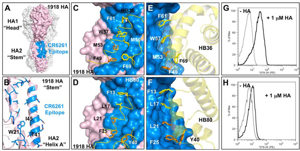
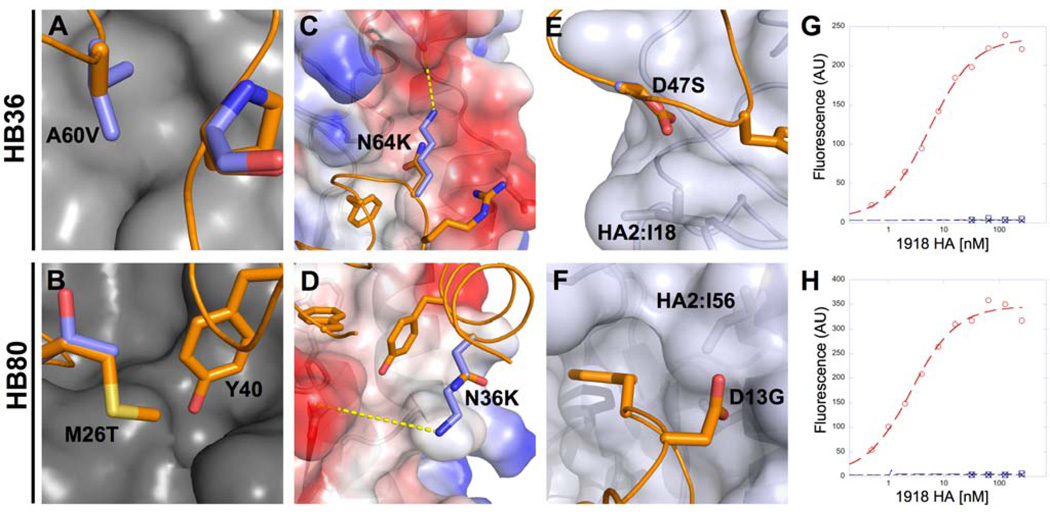
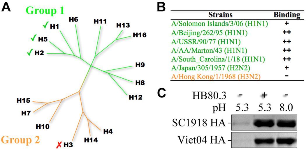
We describe a general computational method for designing proteins that bind a surface patch of interest on a target macromolecule. Favorable interactions between disembodied amino acid residues and the target surface are identified and used to anchor de novo designed interfaces. The method was used to design proteins that bind a conserved surface patch on the stem of the influenza hemagglutinin (HA) from the 1918 H1N1 pandemic virus. After affinity maturation, two of the designed proteins, HB36 and HB80, bind H1 and H5 HAs with low nanomolar affinity. Further, HB80 inhibits the HA fusogenic conformational changes induced at low pH. The crystal structure of HB36 in complex with 1918/H1 HA revealed that the actual binding interface is nearly identical to that in the computational design model. Such designed binding proteins may be useful for both diagnostics and therapeutics.


Figures

Fig. 1
Fig. 1. Overview of the design process
The flow chart illustrates key steps in the design process for novel binding proteins, with thumbnails illustrating each step in the creation of binders that target the stem of the 1918 HA.
Fig. 2
Fig. 2. Design of HB36 and HB80, targeting the stem of the 1918 HA
(A) Surface representation of the trimeric HA structure (PDB 3R2X) from the 1918 pandemic virus, with one of the three protomers highlighted in pink. Broadly neutralizing antibody CR6261 binds a highly conserved epitope in the stem region (blue patch on surface), close to the viral membrane (bottom). (B) Enlarged view of the CR6261 epitope (blue region from part A), with CR6261 contact residues depicted as sticks. This target site on HA contains a groove lined by multiple hydrophobic residues. Loops on either side of this hydrophobic groove (above and below) constrain access to this region. Key residues on HA2 are noted in one-letter code. (C and D) Front view of the designed interaction between HB36 (C) and HB80 (D) and the target site on HA. HA is rotated approximately 60° relative to Fig. 2A. Contact segments of HB36 and HB80 are colored in yellow and residues are depicted as sticks, with hotspot residues highlighted all in orange (F49 and M53 for HB36 and L21, F25, and Y40 for HB80). Polar atoms of side chains are shown in red (oxygen) and blue (nitrogen). For clarity, the non-contacting regions from the designs have been omitted. (E and F) Further details of the designed interactions of HB36 (E) and HB80 (F) with 1918/H1 HA. (G and H) Initial binding data for HB36 (G) and HB80 (H) designs (before affinity maturation). When incubated with 1 µM 1918 HA, yeast displaying the two designed proteins show an increase in fluorescent phycoerythrin signal (x-axis) compared to the absence of 1918 HA. Coordinate files of models of 1918 HA in complex with HB36 and HB80 are available as Supporting Online Material.
Fig. 3
Fig. 3. Affinity Maturation of HB36 and HB80
Substitutions that increase the affinity of the original designs can be classified as deficiencies in modeling the (A and B) repulsive interactions HB36 Ala60Val (A), HB80 Met26Thr (B); (C and D) electrostatics HB36 Asn64Lys (C), HB80 Asn36Lys (D); (E and F) and solvation HB36 Asp47Ser (E), HB80 Asp12Gly (F). Binding titrations of HB36.4 (G) and HB80.3 (H) to SC1918/H1 HA as measured by yeast surface display. Red circles represent the affinity-matured design, blue squares the scaffold protein from which the design is derived, and black crosses represent the design in the presence of 750nM inhibitory CR6261 Fab.
Fig. 4
Fig. 4. Crystal structure of HB36 in complex with SC1918 HA confirms designed interface
(A) Superposition of the crystal structure of HB36.3-SC1918/H1 complex (HB36.3 in red with SC1918 HA1 subunit in pink and HA2 subunit in cyan) and the computational design (blue) reveals good agreement in the position of the main recognition helix, with a slight rotation of the rest of the protein domain. Superposition was performed using the HA2 subunits. For clarity, only the HA from the crystal structure is depicted here (the HA used for superposition of the design, which is essentially identical to the crystal structure, was omitted). (B) Close up of the SC1918 HA-HB36.3 interface, highlighting the close agreement between the design and the crystal structure. The main recognition helix is oriented approximately as in (A), with the HB36.3 crystal structure in red (pink side chains), the design in blue (light blue side chains), and HA in cyan and pink at left. (C) Unbiased 2Fo-Fc (gray mesh, contoured at 1σ) and Fo-Fc (green mesh, contoured at 3σ) electron-density maps for the main recognition helix of HB36.3. The helix is oriented as in (B), with key contact residues of the left face of the helix in this view labeled (the right surface faces and interacts with the core of the HB36.3 protein). Significant density was observed for most of the large side chains at the interface with HA, including F49, M53, W57, F61, and F69 (not visible in this view). While side chains are shown here to illustrate their agreement with the experimental electron density, maps were calculated after initial refinement of an HA-HB36.3 model with the following side chains truncated to alanine (no prior refinement with side chains present): F49, M53, M56, W57, F61, and F69.
Fig. 5
Fig. 5. HB80 binds multiple HA subtypes and inhibits the conformational changes that drive membrane fusion
(A) Phylogenetic tree depicting the relationship between the 16 influenza A hemagglutinin subtypes. These subtypes can be divided into two main lineages, groups 1 and 2. CR6261 has broad activity against group 1 viruses. HB80.3 has a similar cross-reactivity profile and binds multiple group 1 subtypes, including H1 and H5. (B) Binding data for HB80.3 and CR6261 Fab against a panel of HAs. “+”, “++”, and “+++” indicate relative degree of binding (approximately 10−7, 10−8, and 10−9 M, respectively), while “-“ indicates no detectable binding at the highest concentration tested (100nM). (C) HB80.3 inhibits the pH-induced conformational changes that drive membrane fusion. Exposure to low pH converts 1918 H1 HA (top panel) and the Viet04 H5 HA to a protease susceptible state (lane 1), while HAs maintained at neutral pH are highly resistant to trypsin (lane 3). Pre-incubation of HB80.3 with H1 and H5 prevents pH-induced conformational changes and retains the HAs in the protease-resistant, pre-fusion state (lane 2).

Comment in

References

    1. Ledford H. Nature. 2008;455:437. - PubMed
    1. Lerner RA. Angew Chem Int Ed Engl. 2006;45:8106. - PubMed
    1. Kortemme T, et al. Nat. Struct. Mol. Biol. 2004;11:371. - PubMed
    1. Jha RK, et al. J Mol Biol. 2010;400:257. - PMC - PubMed
    1. Huang PS, Love JJ, Mayo SL. Protein Sci. 2007;16:2770. - PMC - PubMed

Publication types

MeSH terms

Substances

Associated data