

Microparticles in hemostasis and thrombosis
- PMID: 21566224
- PMCID: PMC3144708
- DOI: 10.1161/CIRCRESAHA.110.233056
Microparticles in hemostasis and thrombosis
Abstract
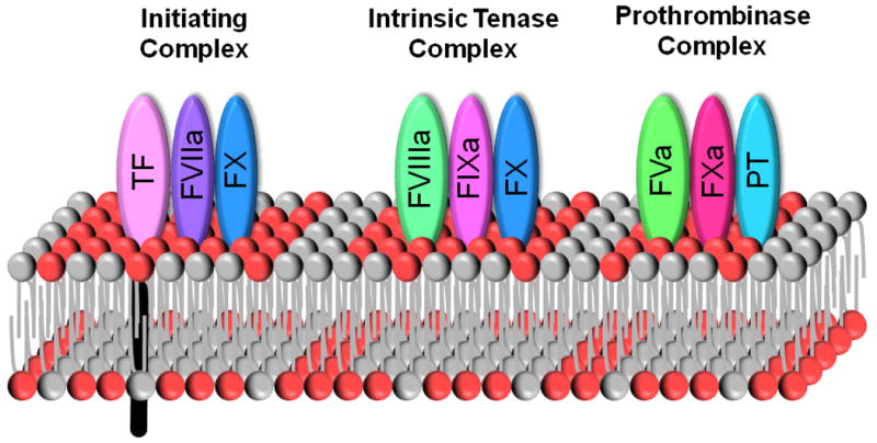
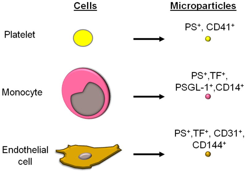
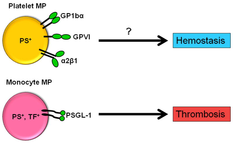
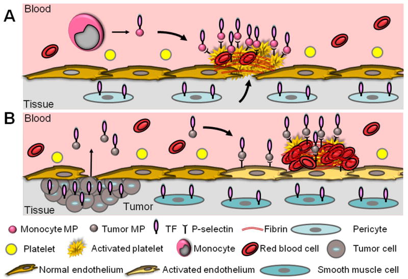
Blood contains microparticles (MPs) derived from a variety of cell types, including platelets, monocytes, and endothelial cells. In addition, tumors release MPs into the circulation. MPs are formed from membrane blebs that are released from the cell surface by proteolytic cleavage of the cytoskeleton. All MPs are procoagulant because they provide a membrane surface for the assembly of components of the coagulation protease cascade. Importantly, procoagulant activity is increased by the presence of anionic phospholipids, particularly phosphatidylserine (PS), and the procoagulant protein tissue factor (TF), which is the major cellular activator of the clotting cascade. High levels of platelet-derived PS(+) MPs are present in healthy individuals, whereas the number of TF(+), PS(+) MPs is undetectable or very low. However, levels of PS(+), TF(+) MPs are readily detected in a variety of diseases, and monocytes appear to be the primary cellular source. In cancer, PS(+), TF(+) MPs are derived from tumors and may serve as a useful biomarker to identify patients at risk for venous thrombosis. This review will summarize our current knowledge of the role of procoagulant MPs in hemostasis and thrombosis.
Figures
References
-
- Burnier L, Fontana P, Kwak BR, Angelillo-Scherrer A. Cell-derived microparticles in haemostasis and vascular medicine. Thromb Haemost. 2009;101:439–451. - PubMed
-
- Morel O, Toti F, Hugel B, Bakouboula B, Camoin-Jau L, Dignat-George F, Freyssinet JM. Procoagulant microparticles: Disrupting the vascular homeostasis equation? Arterioscler Thromb Vasc Biol. 2006;26:2594–2604. - PubMed
-
- Nieuwland R, Sturk A. Why do cells release vesicles? Thromb Res. 2010;125 1:S49–51. - PubMed
-
- Mause SF, Weber C. Microparticles: Protagonists of a novel communication network for intercellular information exchange. Circ Res. 2010;107:1047–1057. - PubMed
-
- Chargaff E, West R. The biological significance of the thromboplastic protein of blood. J Biol Chem. 1946;166:189–197. - PubMed
Publication types
MeSH terms
Substances
Grants and funding
LinkOut - more resources
Full Text Sources
Other Literature Sources
Medical
Miscellaneous
