

Oral inflammatory myofibroblastic tumor: case report and review of literature
- PMID: 21566695
- PMCID: PMC3091292
- DOI: 10.2174/1874210601105010066
Oral inflammatory myofibroblastic tumor: case report and review of literature
Abstract
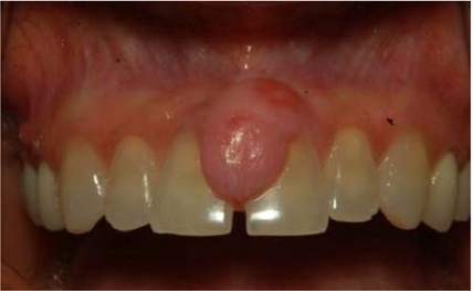
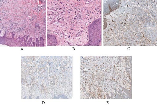
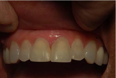
Inflammatory myofibroblastic tumor (IMT) is a rarely described tumor of unknown etiology and pathogenesis. It occurs primarily in the lungs, but has occurred in other extra-pulmonary sites. Histologically these lesions appear as an inflammatory infiltrate within a variably myofibrotic background. Current evidence shows that inflammatory myofibroblastic tumors are neoplastic processes resulting from chromosomal translocations that often cause an overexpression of ALK kinase, which is often assessed using immunohistochemical studies. Currently, the biological behavior of oral inflammatory myofibroblastic tumor is still uncertain. This article describes the clinical, histological, and operative features of a case of IMT of the oral cavity.
Keywords: Inflammatory myofibroblastic tumor; inflammatory pseudotumor; oral cavity..
Figures
References
-
- Narla LD, Newman B, Spottswood SS, Narla S, Koll IR. Inflammatory pseudotumor. Radiographics. 2003;23:719–29. - PubMed
-
- Van Weert S, Manni JJ, Driessen A. Inflammatory myofibroblastic tumor of the parotid gland: case report and review of the literature. Acta Otolaryngol. 2005;125:433–7. - PubMed
-
- Ide F, Shimoyama T, Horie N. Intravenous myofibroblastic pseudotumour of the buccal mucosa. Oral Oncol. 1998;34:232–5. - PubMed
-
- Liston SL, Dehner LP, Jarvis CW, et al. Inflammatory pseudotumors in the buccal tissues of children. Oral Surg Oral Med Oral Pathol. 1981;51:287–91. - PubMed
LinkOut - more resources
Full Text Sources