

Right ventrolateral prefrontal cortex mediates individual differences in conflict-driven cognitive control
- PMID: 21568631
- PMCID: PMC3641154
- DOI: 10.1162/jocn_a_00064
Right ventrolateral prefrontal cortex mediates individual differences in conflict-driven cognitive control
Abstract
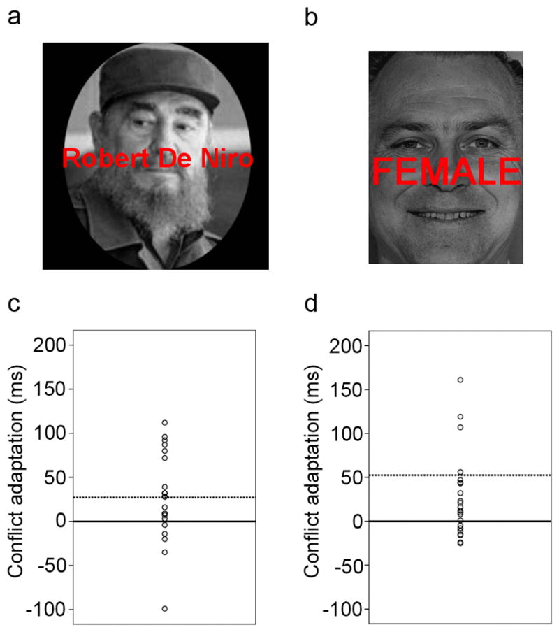
Conflict adaptation--a conflict-triggered improvement in the resolution of conflicting stimulus or response representations--has become a widely used probe of cognitive control processes in both healthy and clinical populations. Previous fMRI studies have localized activation foci associated with conflict resolution to dorsolateral PFC (dlPFC). The traditional group analysis approach employed in these studies highlights regions that are, on average, activated during conflict resolution, but does not necessarily reveal areas mediating individual differences in conflict resolution, because between-subject variance is treated as noise. Here, we employed a complementary approach to elucidate the neural bases of variability in the proficiency of conflict-driven cognitive control. We analyzed two independent fMRI data sets of face-word Stroop tasks by using individual variability in the behavioral expression of conflict adaptation as the metric against which brain activation was regressed while controlling for individual differences in mean RT and Stroop interference. Across the two experiments, a replicable neural substrate of individual variation in conflict adaptation was found in ventrolateral PFC (vlPFC), specifically, in the right inferior frontal gyrus, pars orbitalis (BA 47). Unbiased regression estimates showed that variability in activity in this region accounted for ∼ 40% of the variance in behavioral expression of conflict adaptation across subjects, thus documenting a heretofore unsuspected key role for vlPFC in mediating conflict-driven adjustments in cognitive control. We speculate that vlPFC plays a primary role in conflict control that is supplemented by dlPFC recruitment under conditions of suboptimal performance.
Figures


References
-
- Aron AR. The neural basis of inhibition in cognitive control. Neuroscientist. 2007;13(3):214–228. - PubMed
-
- Aron AR, Robbins TW, Poldrack RA. Inhibition and the right inferior frontal cortex. Trends Cogn Sci. 2004;8(4):170–177. - PubMed
-
- Barbas H. Anatomic organization of basoventral and mediodorsal visual recipient prefrontal regions in the rhesus monkey. J Comp Neurol. 1988;276(3):313–342. - PubMed
-
- Botvinick MM, Braver TS, Barch DM, Carter CS, Cohen JD. Conflict monitoring and cognitive control. Psychol Rev. 2001;108(3):624–652. - PubMed
Publication types
MeSH terms
Grants and funding
LinkOut - more resources
Full Text Sources
Miscellaneous