Review
. 2011 Aug 15:13:345-68.
doi: 10.1146/annurev-bioeng-071910-124654.

Mapping fetal brain development in utero using magnetic resonance imaging: the Big Bang of brain mapping

Affiliations
Review

Mapping fetal brain development in utero using magnetic resonance imaging: the Big Bang of brain mapping

Colin Studholme. Annu Rev Biomed Eng. .

Abstract

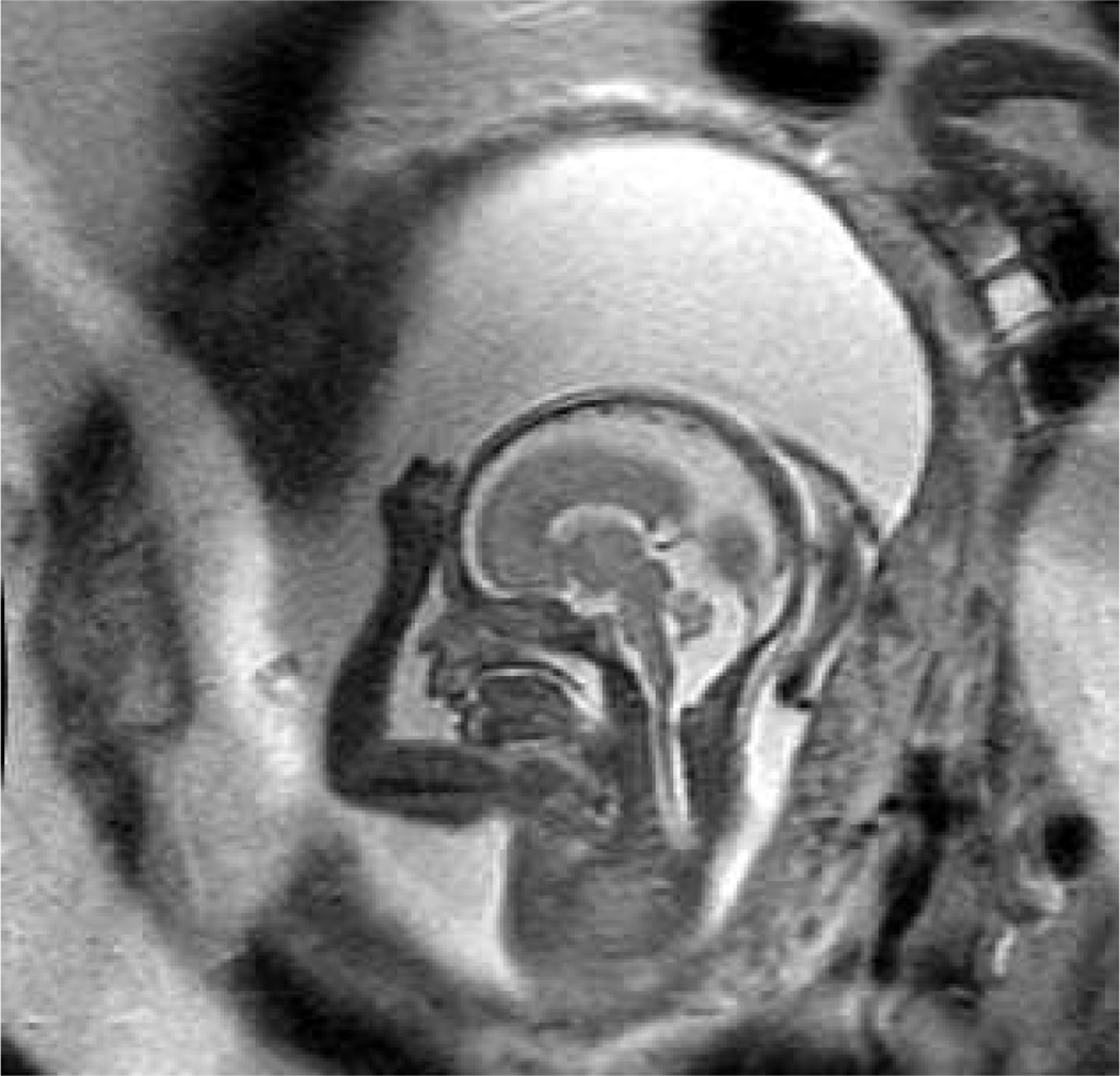
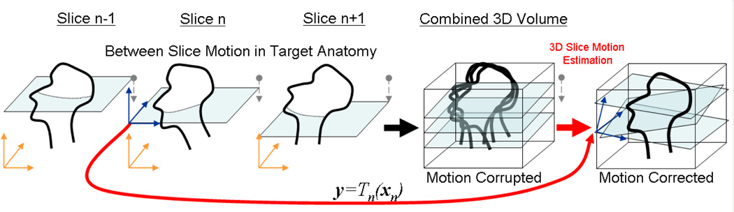
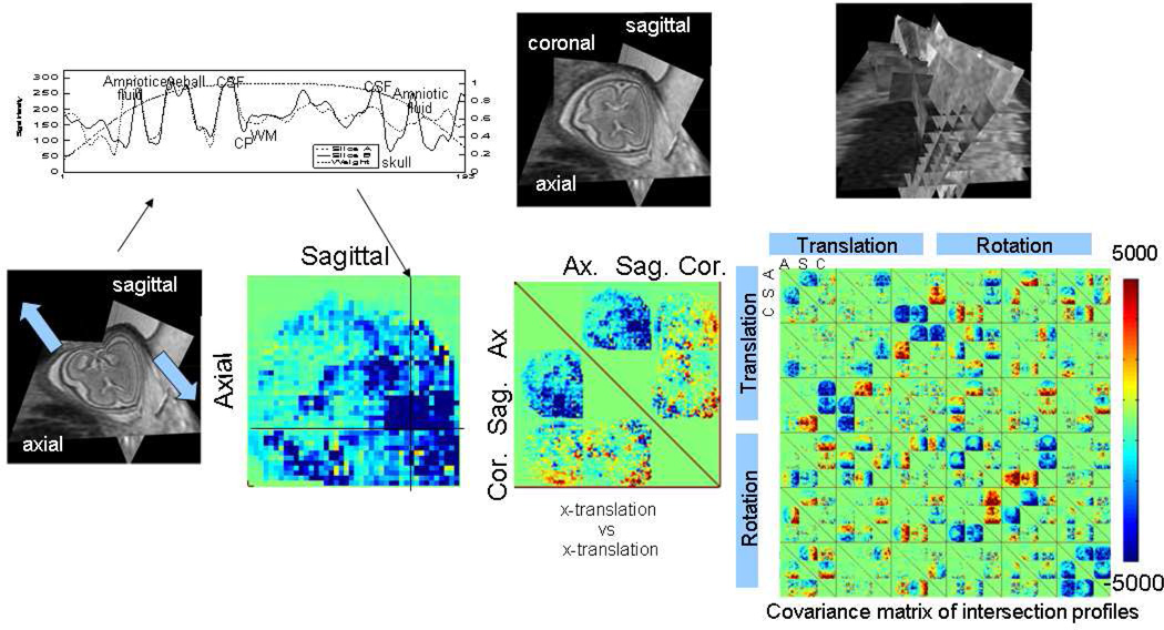
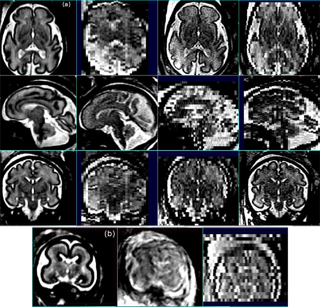
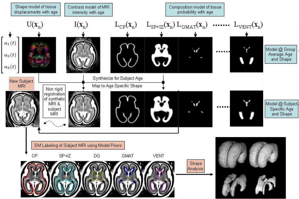
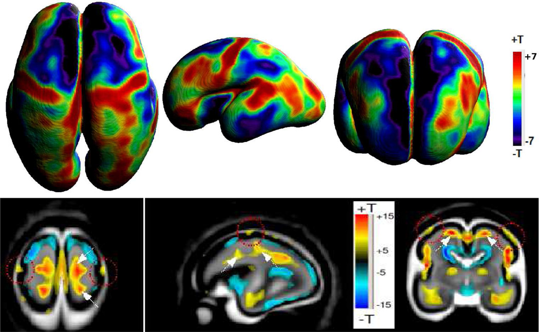
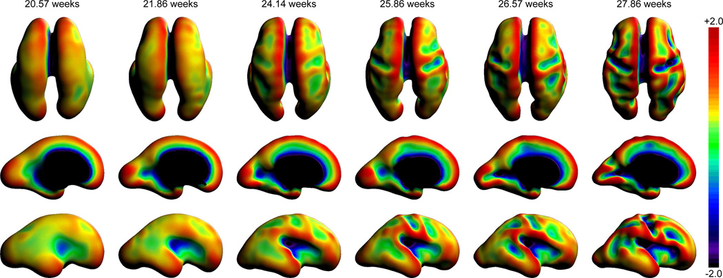
The development of tools to construct and investigate probabilistic maps of the adult human brain from magnetic resonance imaging (MRI) has led to advances in both basic neuroscience and clinical diagnosis. These tools are increasingly being applied to brain development in adolescence and childhood, and even to neonatal and premature neonatal imaging. Even earlier in development, parallel advances in clinical fetal MRI have led to its growing use as a tool in challenging medical conditions. This has motivated new engineering developments encompassing optimal fast MRI scans and techniques derived from computer vision, the combination of which allows full 3D imaging of the moving fetal brain in utero without sedation. These promise to provide a new and unprecedented window into early human brain growth. This article reviews the developments that have led us to this point, examines the current state of the art in the fields of fast fetal imaging and motion correction, and describes the tools to analyze dynamically changing fetal brain structure. New methods to deal with developmental tissue segmentation and the construction of spatiotemporal atlases are examined, together with techniques to map fetal brain growth patterns.


Figures

Figure 1
Figure 1
Example slice from a clinical fast (SSFSE) T2W MRI image of a human fetus showing a mid-line sagittal view of the fetal brain.
Figure 2
Figure 2
The motion scattered multislice to volume matching problem: Combining fast multislice MRI acquisitions by retrospective fusion to a 3D volume: Each individual slice n making up the volume must be accurately located and orientated within the final 3D anatomical space by a full 3D rigid transformation Tn(xn) consisting of 3 rotations and 3 translations. Once these are estimated a final volume image can be reconstructed on a regular voxel lattice using scattered data interpolation techniques.
Figure 3
Figure 3
An illustration of intersection based slice motion estimation: Any intersecting pairs of slices through the moving fetal head must match along their intersection when mapped correctly together into the 3D anatomical space. The square difference in intensity along an intersection is related to corresponding changes in the spatial transformation applied to each slice. By considering all slice pairs acquired in a multislice study consisting of many stacks of slices, a numerical optimization scheme can be formulated to minimize discrepancies, using the covariance matrix of all matches with respect to the slice transformation parameters. This collectively adjusts the 3 translations and 3 rotations of each slice to bring them into mutual alignment.
Figure 4
Figure 4
(a) Example motion estimation (107), relative intensity bias correction (113) and reconstruction into a single 3D volume (orthogonal slices in left column) of individual sagittal, axial and coronal slice stacks (right three columns) acquired from a clinical fetal brain study using fast multi-slice imaging (note the original slices are shown in acquired coordinates before motion estimation and therefore are not precisely aligned). These illustrate the valuable increase (left column) in the isotropic resolution and contrast to noise resulting from the accurate fusion of multiple fast multi-slice imaging studies of the moving fetus. (b) Motion correction and fusion of MRI scans of a younger fetal brain with extreme motion shown: reconstructed with individual slice motion estimation (left) and without (center), together one of the original slice stacks (right).
Figure 5
Figure 5
An illustration of automated atlas based tissue segmentation of a 3D T2W fetal brain MRI using an age specific prior map of tissue probability, MRI contrast and brain shape and size. Labels assigned to each voxel dividing the brain into Cortical Plate, Intermediate Zone and Sub-Plate, Deep Grey Matter, Germinal Matrix and Ventricular CSF, are adapted from the atlas prior to fit the subject MRI scan using an iterative Expectation Maximization algorithm (132).
Figure 6
Figure 6
Example Tensor Based Morphometry of the pattern of tissue growth between 20 and 28 gestational weeks: Statistical significance maps show points where the rate of volume increase is greater or lesser than the rate of growth of the brain as a whole. The lower row shows slices within the brain volume, while the top row shows differences across the cortical plate only. These local variations in tissue growth rate create structural complexity as the primary sulci form.
Figure 7
Figure 7
Example maps of surface mean curvature of the boundary between the cortical plate and the sup-plate (equivalent to the gray matter/white matter interface in adults), evaluated on surface mesh representations automatically constructed from in utero MRI scans of fetuses with different gestational ages. These illustrate surface folding progression in terms of mean curvature increases (convexity) and decreases (concavity) as the early primary sulci common to all adults form. These include the sylvian fissure, the pre and post central sulci and the calcarine sulcus.

Similar articles

Cited by

References

    1. Toga Arthur W, Thompson Paul M. Temporal dynamics of brain anatomy. Annual Review of Biomedical Engineering. 2003;5:119–145. - PubMed
    1. Mazziotta JC, Toga AW, Evans AC, Fox P, Lancaster J. A probabilistic atlas of the human brain: theory and rationale for its development. NeuroImage. 1995;2:89–101. - PubMed
    1. Miller Michael I, Trouve Alain, Younes Laurent. On the metrics and euler-lagrange equations of computational anatomy. Annual Review of Biomedical Engineering. 2002;4:375–405. - PubMed
    1. Thompson Paul M, Mega1 Michael S, Woods Roger P, Zoumalan Chris I, Lindshield Chris J, Blanton Rebecca E, Moussai Jacob, Holmes Colin J, Cummings Jeffrey L, Toga Arthur W. Cortical change in alzheimer’s disease detected with a disease-specific population-based brain atlas. Cereb. Cortex. 2001;11(1):1–16. - PubMed
    1. Lerch JP, Pruessner JC, Zijdenbos A, Hampel H, Teipel SJ, Evans AC. Focal decline of cortical thickness in alzheimers disease identified by computational neuroanatomy. Cereb Cortex. 2005;15:995–1001. - PubMed

Publication types

MeSH terms