Review
. 2012 May 1;16(9):869-82.
doi: 10.1089/ars.2011.4019. Epub 2011 Jul 7.

Mitochondrial quality control and dynamics in Parkinson's disease

Affiliations
Review

Mitochondrial quality control and dynamics in Parkinson's disease

Melissa K McCoy et al. Antioxid Redox Signal. .

Abstract

Significance: Studies of sporadic cases, toxin models, and genetic causes of Parkinson's disease suggest that mitochondrial dysfunction may be an early feature of pathogenesis.

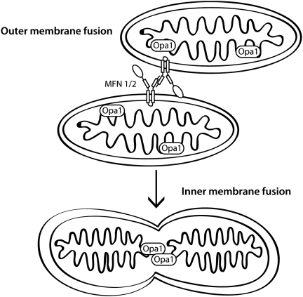
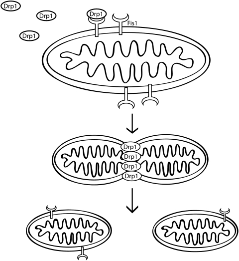
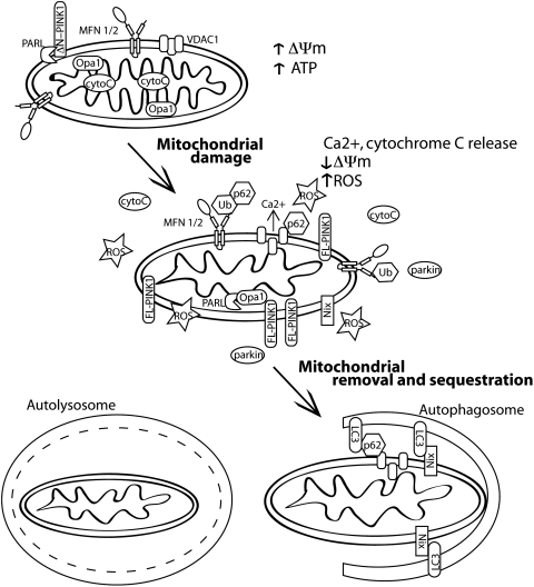
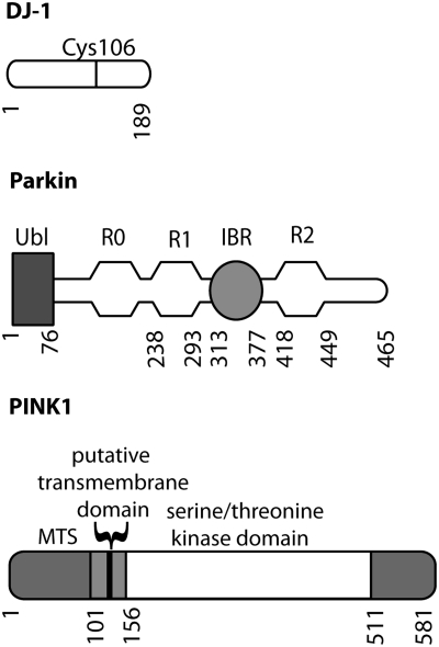
Recent advances: Compelling evidence of a causal relationship between mitochondrial function and disease was found with the identification of several genes for recessive parkinsonism, PINK1, DJ-1, and parkin. There is evidence that each of these regulates responses to cellular stresses, including oxidative stress and depolarization of the mitochondrial membrane. Specifically, PINK1 and parkin modulate mitochondrial dynamics by promoting autophagic removal of depolarized mitochondria. Mutations in all genes linked to Parkinson's disease lead to enhanced sensitivity to mitochondrial toxins and oxidative stress.

Critical issues: Both increased mitochondrial damage due to complex 1 inhibition, mishandling of calcium, oxidant stress, or impaired clearance of dysfunctional mitochondria would lead to the accumulation of nonfunctional organelles and could contribute to neuronal dysfunction. However, several unanswered questions remain about the underlying mechanism(s) involved.

Future directions: PINK1 and parkin have been demonstrated to regulate mitochondrial dynamics, but the pathways linking PINK1 activity to parkin function are still unclear and warrant further investigation.


Figures

FIG. 1.
FIG. 1.
Mitochondrial fusion. Outer mitochondrial membrane fusion requires mitofusins located on the outer mitochondrial membrane by two transmembrane domains that tether adjacent mitochondria together by oligomerization of Mfn molecules located on opposing organelles. Fusion of the outer mitochondrial membranes requires GTPase activity. Following outer membrane fusion, OPA1, an intermembrane protein, mediates fusion of the inner mitochondrial membrane, which requires oligomerization of OPA1 and GTPase activity.
FIG. 2.
FIG. 2.
Mitochondrial fission. Drp1 and Fis1 promote mitochondrial fission, the process by which a single organelle is cleaved into two or more daughter mitochondria. Drp1 is usually present in the cytoplasm as oligomers, but is recruited to the outer mitochondrial membranes by interaction with FIS1. Once at the membrane, oligomers assemble into larger complexes where they promote GTPase dependent remodeling and fission of the membrane.
FIG. 3.
FIG. 3.
Regulation of mammalian mitophagy. When mitochondria become damaged, they are characterized by reduced membrane potential, they release calcium and cytochrome C and generate reactive oxygen species (ROS). Nonfunctional mitochondria are then removed by mitophagy to alleviate ROS and cytochrome C release into the cytosol and prevent apoptosis. There are several identified pathways to mitophagy activation. Nix is an outer mitochondrial membrane protein that binds to LC3 to promote mitochondrial engulfment by autophagic membranes. Another pathway induced by mitochondrial depolarization occurs when full length PINK1 (FL-PINK1) is no longer proteolysed and accumulates on the outer mitochondrial membrane. Increased levels of membrane-associated PINK1 recruit parkin to the mitochondria. Parkin ubiquitinates mitochondrial substrates, including VDAC and MFN. These ubiquitin modified substrates are recognized by the adaptor protein p62 and are trafficked to autophagosomes and eventually are degraded in lysosomes.
FIG. 4.
FIG. 4.
Genes associated with autosomal recessive early onset parkinsonism. Diagrams of the domain organization of recessive mutations are shown. DJ-1 is a small single domain protein. PINK1 contains a mitochondrial targeting sequence (MTS), a putative transmembrane (TM) region to anchor PINK1 in the outer mitochondrial membrane, and a serine/threonine kinase domain. Parkin has an N-terminal ubiquitin-like (Ubl) domain, followed by three RING finger domains with an in-between RING (IBR) domain after the first two RING finger domains.
FIG. 5.
FIG. 5.
Regulation of mitochondrial transport by PINK1. Mitochondria are transported along microtubules by kinesin motors for distribution throughout the cytosol. Both Mfn2 and full-length (FL) or N-terminally cleaved (ΔN) PINK 1 bind the Miro/Milton complex that connects mitochondria to kinesin and promote axonal transport of mitochondria along microtubules.
FIG. 6.
FIG. 6.
Regulation of mitophagy by PINK1. PINK1-dependent mitophagy requires the accumulation of full-length (FL) PINK1 on the outer mitochondrial membrane that recruits parkin to mediate selective mitophagy through increased ubiquitination of substrates, including Mfn2. PINK1 and can also bind beclin1 to promote autophagy through the class III PI3 kinase VPS34 and p150.
FIG. 7.
FIG. 7.
Genes associated with autosomal dominant parkinsonism. Diagrams of the domain organization of dominant mutations not drawn to scale are shown. LRRK2 is a large multidomain protein. The N-terminal region contains Ankyrin (ANK) and Leucine-rich repeats (LRR). Following the repeats is the core catalytic region of the protein containing a GTP-binding Ras of complex protein (Roc) domain, a carboxy-terminal of Roc (COR) domain, and a kinase domain. Following the kinase domain is a C-terminal WD40 domain ending before a short C-terminal tail. The N-terminal region of α-synuclein is important for membrane association and is composed of imperfect repeats of KTEGV. The next region of α-synuclein is hydrophobic NAC (nonamyloid component) region, followed by an unstructured C-terminal acidic tail.

References

    1. Ahlqvist G. Landin S. Wroblewski R. Ultrastructure of skeletal muscle in patients with Parkinson's disease and upper motor lesions. Lab Invest. 1975;32:673–679. - PubMed
    1. Alexander C. Votruba M. Pesch UE. Thiselton DL. Mayer S. Moore A. Rodriguez M. Kellner U. Leo-Kottler B. Auburger G. Bhattacharya SS. Wissinger B. OPA1, encoding a dynamin-related GTPase, is mutated in autosomal dominant optic atrophy linked to chromosome 3q28. Nat Genet. 2000;26:211–215. - PubMed
    1. Alirol E. James D. Huber D. Marchetto A. Vergani L. Martinou JC. Scorrano L. The mitochondrial fission protein hFis1 requires the endoplasmic reticulum gateway to induce apoptosis. Mol Biol Cell. 2006;17:4593–4605. - PMC - PubMed
    1. Andreyev AY. Kushnareva YE. Starkov AA. Mitochondrial metabolism of reactive oxygen species. Biochemistry (Mosc) 2005;70:200–214. - PubMed
    1. Anglade P. Vyas S. Javoy-Agid F. Herrero MT. Michel PP. Marquez J. Mouatt-Prigent A. Ruberg M. Hirsch EC. Agid Y. Apoptosis and autophagy in nigral neurons of patients with Parkinson's disease. Histol Histopathol. 1997;12:25–31. - PubMed

Publication types