. 2011 May 10:7:32.
doi: 10.1186/1744-8069-7-32.

Nav1.7 is the predominant sodium channel in rodent olfactory sensory neurons

Affiliations

Nav1.7 is the predominant sodium channel in rodent olfactory sensory neurons

Hye-Sook Ahn et al. Mol Pain. .

Abstract

Background: Voltage-gated sodium channel Nav1.7 is preferentially expressed in dorsal root ganglion (DRG) and sympathetic neurons within the peripheral nervous system. Homozygous or compound heterozygous loss-of-function mutations in SCN9A, the gene which encodes Nav1.7, cause congenital insensitivity to pain (CIP) accompanied by anosmia. Global knock-out of Nav1.7 in mice is neonatal lethal reportedly from starvation, suggesting anosmia. These findings led us to hypothesize that Nav1.7 is the main sodium channel in the peripheral olfactory sensory neurons (OSN, also known as olfactory receptor neurons).

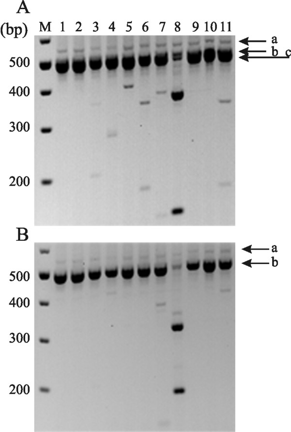
Methods: We used multiplex PCR-restriction enzyme polymorphism, in situ hybridization and immunohistochemistry to determine the identity of sodium channels in rodent OSNs.

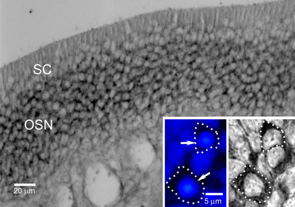
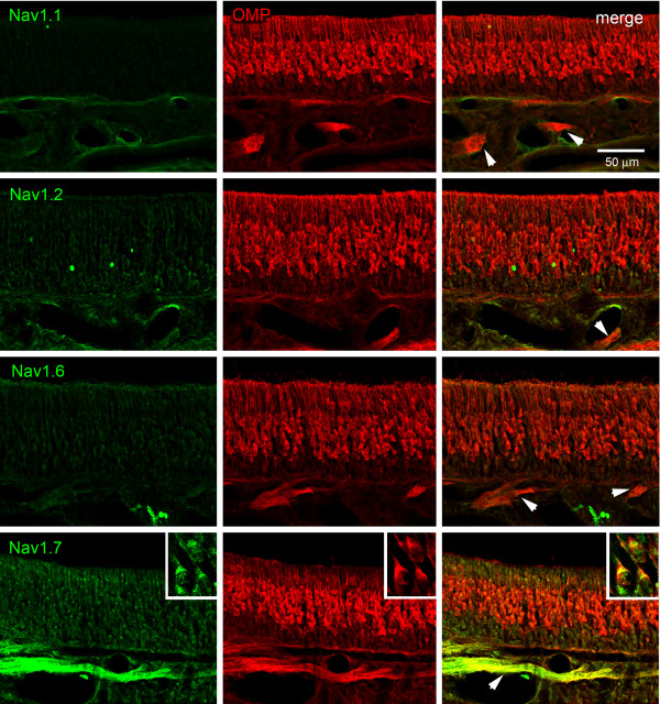
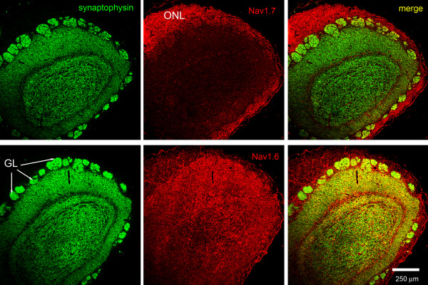
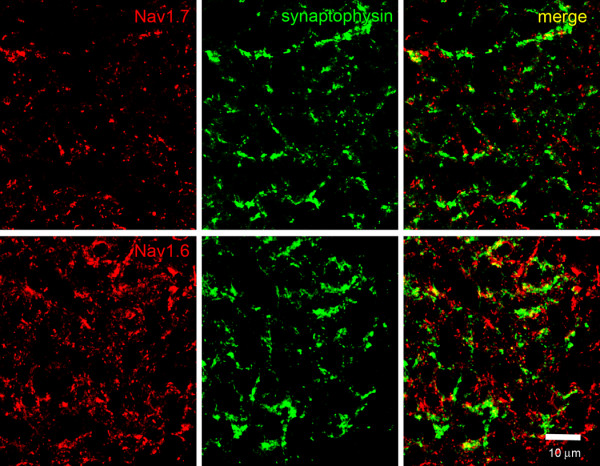
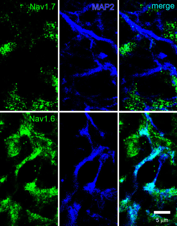
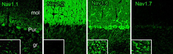
Results: We show here that Nav1.7 is the predominant sodium channel transcript, with low abundance of other sodium channel transcripts, in olfactory epithelium from rat and mouse. Our in situ hybridization data show that Nav1.7 transcripts are present in rat OSNs. Immunostaining of Nav1.7 and Nav1.6 channels in rat shows a complementary accumulation pattern with Nav1.7 in peripheral presynaptic OSN axons, and Nav1.6 primarily in postsynaptic cells and their dendrites in the glomeruli of the olfactory bulb within the central nervous system.

Conclusions: Our data show that Nav1.7 is the dominant sodium channel in rat and mouse OSN, and may explain anosmia in Nav1.7 null mouse and patients with Nav1.7-related CIP.


Figures

Figure 1
Figure 1
Restriction analysis of multiplex PCR amplicons of sodium channels from adult rat and mouse olfactory epithelium. (A) Lane M shows 100-bp ladder marker, and lane 1 contains amplicon from rat olfactory epithelium. Bands "a", "b" and "c" (best seen in Lane 8) are consistent with the presence of a potential mixture of sodium channels. Band "a": Nav1.1 (558 bp), Nav1.2 (561 bp) and Nav1.3 (561 bp), band "b": Nav1.5 (519 bp); band "c": Nav1.6 (507 bp), Nav1.7 (501 bp), Nav1.8 (480 bp), Nav1.9 (468 bp) and Nax (501 bp). Lanes 2-11 show results of cutting this DNA with EcoRV (Nav1.1), EcoNI (Nav1.2), AvaI (Nav1.3), NaeI (Nav1.4/Nax), AccI (Nav1.5/1.9), SphI (Nav1.6), BamHI (Nav1.7/1.8), AflII (Nav1.8), EcoRI (Nav1.9), and XbaI (Nax). (B) Lane M shows 100-bp ladder marker, and lane 1 contains amplicons from mouse olfactory epithelium. Bands "a" and "b" are consistent with the presence of a potential mixture of sodium channels. Band "a": Nav1.1 (558 bp), Nav1.2 (561 bp) and Nav1.3 (561 bp); band "b": Nav1.6 (510 bp), Nav1.7 (501 bp), Nav1.8 (480 bp), Nav1.9 (471 bp) and Nax (501 bp). Lanes 2-11 show the results of cutting this DNA with EcoRV (Nav1.1), EcoNI (Nav1.2), DraI (Nav1.3), PvuI (Nav1.4), AgeI (Nav1.5), SphI (Nav1.6), ScaI (Nav1.7), AhdI (Nav1.8), EcoRI (, Nav1.9) and AlwNI (Nax).
Figure 2
Figure 2
Nav1.7 mRNA expression in olfactory epithelium using in situ hybridization. In situ hybridization signal is exhibited by olfactory sensory neurons (OSN) in the olfactory epithelium. Sustentacular cells (SC) do not express Nav1.7 mRNA signal above background levels. Inset: Increased magnification demonstrates Nav1.7 in situ hybridization signal in the peri-nuclear cytoplasm of OSN. The cell boundaries of two OSN that exhibit robust Nav1.7 signal are demarcated by dotted lines; the DAPI-labeled nuclei of these cells are indicated by arrows.
Figure 3
Figure 3
Sodium channel protein expression in rat olfactory epithelium. Immunostaining experiments using antibodies specific for sodium channels Nav1.1, Nav1.2 and Nav1.6 show that these channels are not detected within olfactory epithelium or in the subjacent olfactory nerve branches (arrows). In contrast, Nav1.7 immunolabeling is displayed in the olfactory epithelium and is robustly expressed within branches of the olfactory nerve (arrows). Insets: At increased magnification, Nav1.7 immunoreactivity (green) is displayed by OMP-positive OSN.
Figure 4
Figure 4
Sodium channels Nav1.6 and Nav1.7 expression in olfactory nerve layer of rat olfactory bulb. Immunolabeling experiments show that sodium channel Nav1.7 (red) is co-localized (yellow) with peripherin (green) in fibers of the olfactory nerve layer (ONL). In contrast, only limited Nav1.6 immunoreactivity is displayed within the olfactory nerve layer, with robust labeling of the glomeuli (GL).
Figure 5
Figure 5
Sodium channels Nav1.6 and Nav1.7 expression in rat glomerular layer of olfactory bulb. Immunolabeling experiments show that the synaptophysin-positive (green) glomeruli of the olfactory bulb exhibit extremely limited Nav1.7 (red) immunoreactivity. In contrast, Nav1.6 (red) exhibits robust immunoreactivity within the synptophysin-positive (green) glomeruli of the olfactory bulb.
Figure 6
Figure 6
Sodium channels Nav1.6 and Nav1.7 expression in terminal boutons of rat glomerular layer of olfactory bulb. Nav1.7 (red) immunoreactivity within the olfactory bulb glomerulus is punctate and extremely limited, and only occasional co-localization of Nav1.7 with synaptophysin-positive (green) terminal boutons is observed. In contrast, there is robust Nav1.6 (red) immunoreactivity within the glomerulus, although there is limited co-localization with synaptophysin-positive (green) terminal boutons within the glomerulus of the olfactory bulb.
Figure 7
Figure 7
Sodium channel expression within glomerulus dendrites in rat olfactory bulb. MAP2-positive (blue) dendrites do not exhibit detectable Nav1.7 (green) immunoreactivity. In contrast, Nav1.6 immunolabeling is displayed within most MAP2-positive dendrites (co-localization is cyan).
Figure 8
Figure 8
Voltage-dependent activation and fast-inactivation for sodium currents in mouse OSN. (A), sodium currents were evoked by 100 ms depolarizing pulses from -90 mV to +50 mV in steps of 5 mV every 5 s from a holding potential of -100 mV. (B), Normalized peak current-voltage relationship. (C), The inward current was completely blocked by 300 nM TTX. (D), Voltage dependence of activation and steady-state fast-inactivation. To measure steady-state fast-inactivation, sodium currents were elicited by 500 ms prepulses from -160 mV to -20 mV stepped by 10 mV and followed by a 40 ms depolarizing pulse to -20 mV from a holding potential of -100 mV. The activation and fast-inactivation curves were obtained from a Boltzmann fit to the normalized conductance.
Figure 9
Figure 9
Sodium channel immunolabeling in rat cerebellum and DRG. Isoform-specific antibodies generated against sodium channels Nav1.1, Nav1.2, Nav1.6 and Nav1.7 were reacted with sections of adult rat cerebellum and DRG. Purkinje cell bodies and apical dendrites exhibit robust Nav1.1 immunolabeling, while limited Nav1.1 immunoreactivity is displayed in DRG neurons (inset). Parallel fibers of cerebellar granule cells exhibit substantial Nav1.2 labeling; Nav1.2 is not detectable in DRG neurons (inset). Nav1.6 is robustly expressed in Purkinje cell bodies and dendrites and is also localized within parallel fibers of cerebellar granule cells; Nav1.6 immunolabeling is exhibited by most neurons within DRG (inset). Within cerebellum, Nav1.7 immunostaining is not detectable, but Nav1.7 immunoreactivity is exhibited by many DRG neurons. The labeling patterns obtained with the isoform-specific sodium channel antibodies Nav1.1, Nav1.2, Nav1.6 and Nav1.7 utilized in these studies is consistent with previous descriptions of their localization within CNS and PNS tissue [4,29].

References

    1. Firestein S. How the olfactory system makes sense of scents. Nature. 2001;413(6852):211–218. - PubMed
    1. Kaupp UB. Olfactory signalling in vertebrates and insects: differences and commonalities. Nat Rev Neurosci. 2010;11(3):188–200. - PubMed
    1. Trombley PQ, Westbrook GL. Voltage-gated currents in identified rat olfactory receptor neurons. J Neurosci. 1991;11(2):435–444. - PMC - PubMed
    1. Dib-Hajj SD, Cummins TR, Black JA, Waxman SG. Sodium Channels in Normal and Pathological Pain. Annu Rev Neurosci. 2010;33:325–347. - PubMed
    1. Djouhri L, Newton R, Levinson SR, Berry CM, Carruthers B, Lawson SN. Sensory and electrophysiological properties of guinea-pig sensory neurones expressing Nav1.7 (PN1) Na+ channel alpha-subunit protein. J Physiol (Lond) 2003;546(Pt 2):565–576. - PMC - PubMed

Publication types

MeSH terms

LinkOut - more resources