Comparative Study
. 2011 May 10:11:23.
doi: 10.1186/1472-6807-11-23.

Crystal structures of a halophilic archaeal malate synthase from Haloferax volcanii and comparisons with isoforms A and G

Affiliations
Comparative Study

Crystal structures of a halophilic archaeal malate synthase from Haloferax volcanii and comparisons with isoforms A and G

Colten D Bracken et al. BMC Struct Biol. .

Abstract

Background: Malate synthase, one of the two enzymes unique to the glyoxylate cycle, is found in all three domains of life, and is crucial to the utilization of two-carbon compounds for net biosynthetic pathways such as gluconeogenesis. In addition to the main isoforms A and G, so named because of their differential expression in E. coli grown on either acetate or glycolate respectively, a third distinct isoform has been identified. These three isoforms differ considerably in size and sequence conservation. The A isoform (MSA) comprises ~530 residues, the G isoform (MSG) is ~730 residues, and this third isoform (MSH-halophilic) is ~430 residues in length. Both isoforms A and G have been structurally characterized in detail, but no structures have been reported for the H isoform which has been found thus far only in members of the halophilic Archaea.

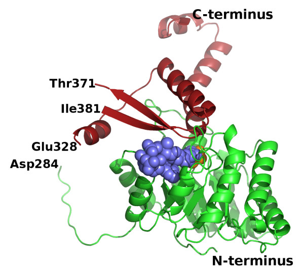
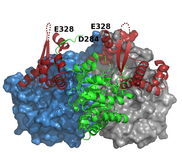
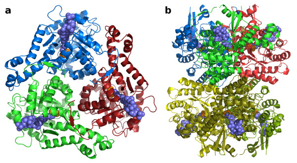
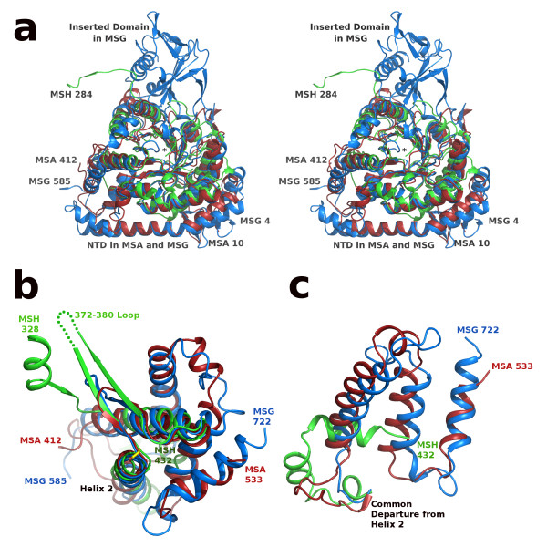
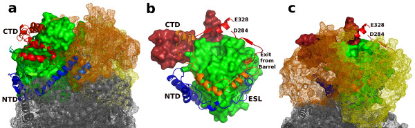
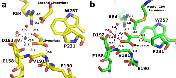
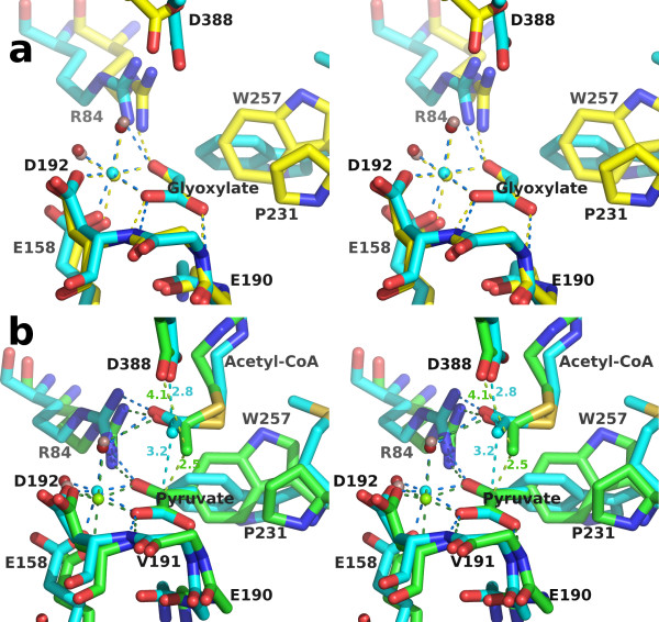
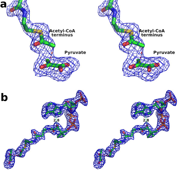
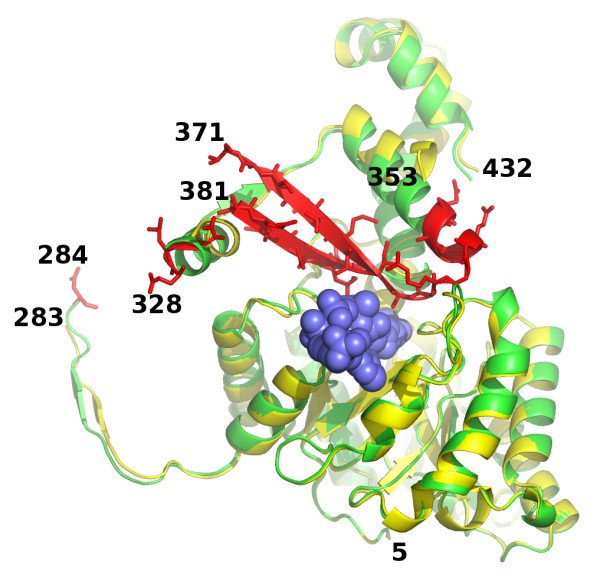
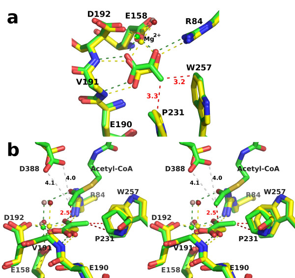
Results: We have solved the structure of a malate synthase H (MSH) isoform member from Haloferax volcanii in complex with glyoxylate at 2.51 Å resolution, and also as a ternary complex with acetyl-coenzyme A and pyruvate at 1.95 Å. Like the A and G isoforms, MSH is based on a β8/α8 (TIM) barrel. Unlike previously solved malate synthase structures which are all monomeric, this enzyme is found in the native state as a trimer/hexamer equilibrium. Compared to isoforms A and G, MSH displays deletion of an N-terminal domain and a smaller deletion at the C-terminus. The MSH active site is closely superimposable with those of MSA and MSG, with the ternary complex indicating a nucleophilic attack on pyruvate by the enolate intermediate of acetyl-coenzyme A.

Conclusions: The reported structures of MSH from Haloferax volcanii allow a detailed analysis and comparison with previously solved structures of isoforms A and G. These structural comparisons provide insight into evolutionary relationships among these isoforms, and also indicate that despite the size and sequence variation, and the truncated C-terminal domain of the H isoform, the catalytic mechanism is conserved. Sequence analysis in light of the structure indicates that additional members of isoform H likely exist in the databases but have been misannotated.


Figures

Figure 1
Figure 1
Overall fold of an H. volcanii MSH monomer in the ternary complex. The N-terminal β8/α8 barrel and the C-terminal domain are shown as cartoon ribbon traces in green and red respectively. Acetyl-coenzyme A and pyruvate are shown as space-filling models in slate blue and orange respectively. The protein segments between Asp284 and Glu328, and between Thr371 and Ile381 were not visible in the crystal structure and have not been included in the model.
Figure 2
Figure 2
Possible domain swapping in H. volcanii MSH. The subunit of the trimer closest to the viewer is rendered in the same form and colors as in figure 1, with the C-terminal domain on the right. The C-terminal domain on the left, also rendered as a red cartoon ribbon trace would instead be connected to the green β8/α8 barrel if domain swapping occurs. In the domain swap the C-terminal domain connected to the barrel of one subunit (green) would be donated to complete the active site of the neighboring barrel shown as a blue surface rather than its own. The short disordered surface loops (372-380) are depicted as red dotted lines.
Figure 3
Figure 3
H. volcanii MSH oligomerization. a) A trimeric assembly rendered as cartoon ribbon traces, viewed along a crystallographic 3-fold rotation axis. Acetyl-coenzyme A and pyruvate are shown as space-filling models in slate blue and orange respectively. b) A hexameric assembly viewed along a crystallographic 2-fold rotation axis, perpendicular to the view in part a. The top trimer is rendered and colored as in part a.
Figure 4
Figure 4
H. volcanii MSH Gel-filtration mobility. a) Gel-filtration elution profile for malate synthase activity and relative absorbance at 280 nm. b) Best-fit linear calibration curve using Bio-Rad gel-filtration standards. Superimposed are the two malate synthase activity peaks, plotted using the elution volume of each and the respective molecular weight of the corresponding hexameric or trimeric assembly.
Figure 5
Figure 5
SSM overlay of MSA [PDB:3CV2], MSG [PDB:1P7T], and MSH [PDB:3OYZ] pyruvate/Acetyl-CoA ternary complexes. a) Stereoview of the N-terminal regions: MSA 10-412, MSG 4-585 and MSH 5-284 rendered in red, blue and green respectively. Ends of protein chains are labeled with residue numbers except MSH residue 5 which is marked by an asterisk at the center of the image. View is from the bottom of the TIM barrel, opposite the active site; C-terminal domains are not shown for clarity. b) Overlay as in part a, but only showing the C-terminal domains of each protein: MSA 412-533, MSG 585-722, and MSH 328-432. The active site base Asp 388 in MSH is shown (center) in stick form with carbon atoms colored yellow and corresponds to the same position as Asp 447 in MSA and Asp 631 in MSG (within 0.7 and 1.0 Å respectively). Disordered loop in MSH is shown as a green dotted line. c) Overlay as in parts a and b, but only showing extreme C-terminal regions: MSA 463-533, MSG 647-722, and MSH 404-432. Helix 2 refers to the second helix of the C-terminal domain which immediately follows the active site aspartate residue as seen in figure 5b.
Figure 6
Figure 6
Relationship of hvMSH oligomerization interfaces to N-terminal clasps and connecting segments in MSA and MSG. Superposition of hvMSH, ecMSA and ecMSG ternary complexes using SSM in Coot. a) N-terminal domain. MSA and MSG are shown as cartoon ribbon traces with N-terminal (NTD) and C-terminal (CTD) domains colored blue and red respectively. Only portions of the CTD which are not homologous to hvMSH are visible. MSH is shown as a green solvent-accessible surface. Other MSH subunits which make up a trimeric assembly are rendered as copper and yellow mesh surfaces; the subunits of the symmetry-related trimer which forms the hexameric assembly are colored grey and shown as mesh surfaces surrounding cartoon ribbon traces. b) Locations of connecting loops; view is from the right side of part a. MSH is shown as a molecular surface with the barrel domain and C-terminal domain colored green and red respectively. Extended surface loops (ESL; blue) connecting the NTD to barrel domains, and loops (orange) connecting barrel domains to the CTD in MSA and MSG are shown as cartoon ribbon traces. The segments of the connection between the barrel domain and CTD of MSH which are visible in electron density maps are depicted as a red cartoon ribbon trace. c) View identical to b, but including other subunits of the trimeric and hexameric hvMSH assembly colored and rendered as in part a.
Figure 7
Figure 7
The active sites of H. volcanii MSH. a) High-occupancy glyoxylate complex rendered in stick form with carbon atoms yellow, oxygen red, and nitrogen blue. The magnesium ion and coordinating water molecules are shown as spheres in purple and red respectively. Distances are shown in angstroms with hydrogen bonds yellow and metal-ligand bonds in black. The side chain of Val 191 has been removed for clarity. b) Pyruvate/Acetyl-CoA ternary complex rendered as in part a, but with carbon atoms in green and sulfur in tan. Additionally, close contacts are indicated with red dashed lines. Side chains for Glu 190 and Val 191 have been omitted for clarity.
Figure 8
Figure 8
Overlays of the H. volcanii MSH active sites with the corresponding E. coli MSG complexes. Overlays were performed by superimposing glyoxylate molecules in part a, or pyruvate molecules in part b, using LSQ in Coot. Residue numbers refer to the H. volcanii sequence. Side chains at positions 190 and 191, and at corresponding positions in ecMSG, have been omitted for clarity. a) Stereoview of high-occupancy glyoxylate complex overlay [PDB:1D8C for ecMSG]. Rendered as in figure 7a, with hvMSH carbon atoms yellow, and ecMSG carbons blue. The magnesium ion and water molecules are colored blue and red respectively in the ecMSG complex, and yellow and lilac in the hvMSH complex. All hydrogen and metal-ligand bonds are colored yellow in hvMSH, and blue in ecMSG. b) Stereoview of pyruvate/Acetyl-CoA ternary complex overlay [PDB:1P7T for ecMSG]. Rendered as in part a, but with carbons, magnesium ions, and bonding interactions in green or blue for hvMSH or ecMSG respectively. Water molecules are lilac for hvMSH and red for ecMSG. Distances are shown in angstroms to show proximity of the acetyl-CoA methyl carbon to either the catalytic base oxygen (Asp 388) or the ketone carbonyl of pyruvate.
Figure 9
Figure 9
Acetyl-CoA/pyruvate omit map showing close contact and the bent conformation of Acetyl-CoA. An Fo-Fc electron density omit map contoured at 3 σ is superimposed on the the final refined model of acetyl-CoA and pyruvate in stick form. Ten rounds of refinement were performed on a model with both molecules removed, prior to map calculation. a) Stereo view of difference density showing the ~2.5 Å close contact (red dashed line). b) View of the map in the region of the active site and acetyl-CoA binding site. An intramolecular hydrogen bond also observed in the ecMSG ternary complex is shown as a green dashed line.
Figure 10
Figure 10
Overall structural comparison of the two H. volcanii complexes. The two models were superimposed using SSM in Coot. The high-occupancy glyoxylate complex is shown in yellow and the corresponding sequences of the ternary complex in green. Portions of the enzyme that become ordered upon acetyl-CoA binding in the ternary complex are colored red and also depicted in stick form. Acetyl-CoA is colored slate blue in space-filling form.
Figure 11
Figure 11
Structural comparison of the active sites of the two H. volcanii complexes. SSM overlay as in figure 10, but showing detail in the region of the active site. The side chain of Val 191 has been omitted for clarity. a) Top view, roughly perpendicular to the glyoxylate molecule. Carbon atoms, magnesium ions, hydrogen and metal-ligand bonds are colored yellow or green in the glyoxylate or ternary complex respectively. Water molecules are depicted as spheres colored red in the glyoxylate and lilac in the ternary complex. Close contacts to the pyruvate methyl group are shown as red dashed lines with distances in angstroms. b) Side view of part a in stereo. Additionally, the close contact between the acetyl methyl carbon of acetyl-CoA and the ketone carbonyl carbon in pyruvate, and the distances between this methyl carbon and the side chain carboxylate oxygens of Asp 388 are shown in red and grey dashed lines respectively.
Figure 12
Figure 12
Strictly conserved residues among all nine homologous sequences in table 2. a) view looking into the active site of the TIM barrel domain, with the C-terminal domain removed for clarity. The surface is shown in space filling form with main chain peptides and strictly conserved residues colored brick red, and side chains of non-conserved residues colored according to atom type with carbon atoms green. The Cα atoms of nonconserved residues are colored green stippled with red. Acetyl-CoA and pyruvate are depicted in stick form showing their binding locations in the ternary complex with carbon atoms colored slate blue or orange respectively. b) same as in part a, but showing C-terminal domain interactions.

References

    1. Kornberg HL, Krebs HA. Synthesis of cell constituents from C2-units by a modified tricarboxylic acid cycle. Nature. 1957;179(4568):988–991. doi: 10.1038/179988a0. - DOI - PubMed
    1. McKinney JD, Honer zu Bentrup K, Munoz-Elias EJ, Miczak A, Chen B, Chan WT, Swenson D, Sacchettini JC, Jacobs WR Jr, Russell DG. Persistence of Mycobacterium tuberculosis in macrophages and mice requires the glyoxylate shunt enzyme isocitrate lyase. Nature. 2000;406(6797):735–738. doi: 10.1038/35021074. - DOI - PubMed
    1. Lorenz MC, Fink GR. The glyoxylate cycle is required for fungal virulence. Nature. 2001;412(6842):83–86. doi: 10.1038/35083594. - DOI - PubMed
    1. Lohman JR, Olson AC, Remington SJ. Atomic resolution structures of Escherichia coli and Bacillus anthracis malate synthase A: Comparison with isoform G and implications for structure-based drug discovery. Protein Science. 2008;17(11):1935–1945. doi: 10.1110/ps.036269.108. - DOI - PMC - PubMed
    1. Smith CV, Sharma V, Sacchettini JC. TB drug discovery: addressing issues of persistence and resistance. Tuberculosis (Edinb) 2004;84(1-2):45–55. doi: 10.1016/j.tube.2003.08.019. - DOI - PubMed

Publication types

LinkOut - more resources