Comparative Study
. 2011 May 10:12:223.
doi: 10.1186/1471-2164-12-223.

The midgut transcriptome of Phlebotomus (Larroussius) perniciosus, a vector of Leishmania infantum: comparison of sugar fed and blood fed sand flies

Affiliations
Comparative Study

The midgut transcriptome of Phlebotomus (Larroussius) perniciosus, a vector of Leishmania infantum: comparison of sugar fed and blood fed sand flies

Anna Dostálová et al. BMC Genomics. .

Abstract

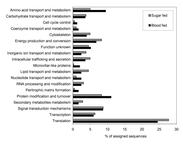
Background: Parasite-vector interactions are fundamental in the transmission of vector-borne diseases such as leishmaniasis. Leishmania development in the vector sand fly is confined to the digestive tract, where sand fly midgut molecules interact with the parasites. In this work we sequenced and analyzed two midgut-specific cDNA libraries from sugar fed and blood fed female Phlebotomus perniciosus and compared the transcript expression profiles.

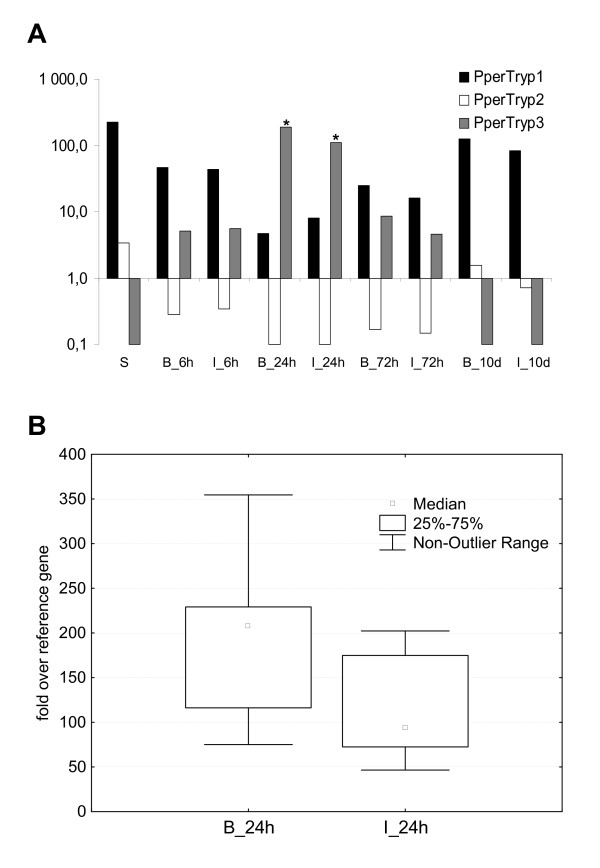
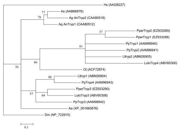
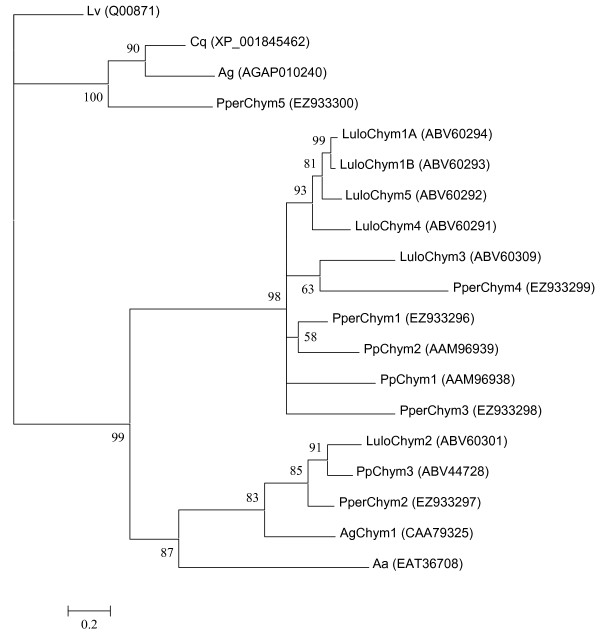
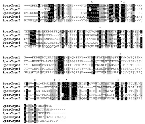
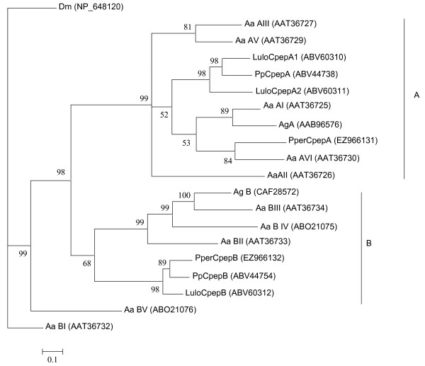
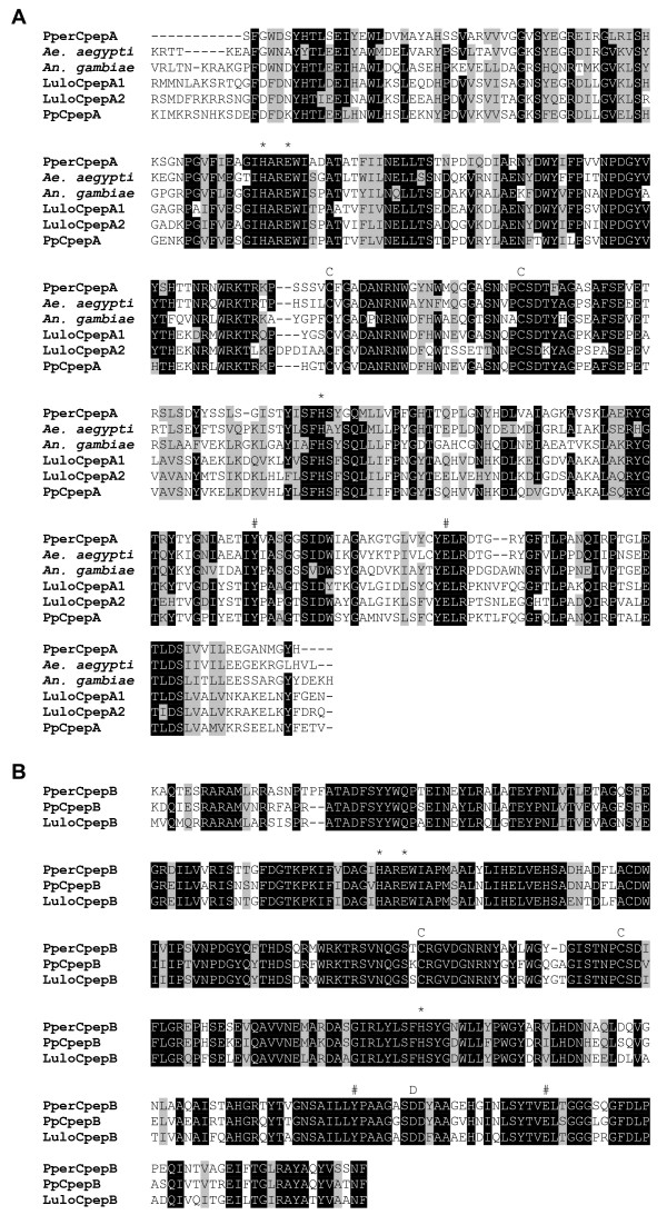
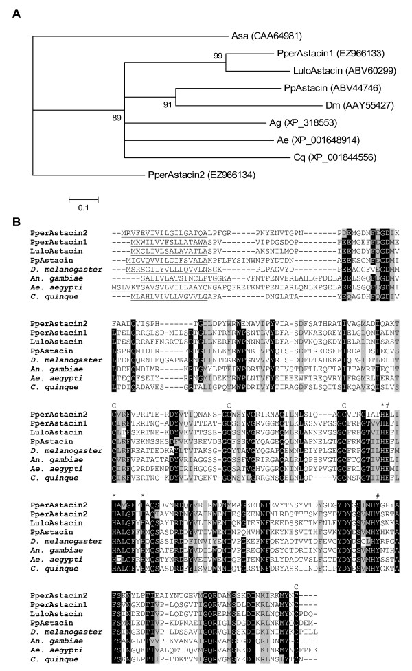
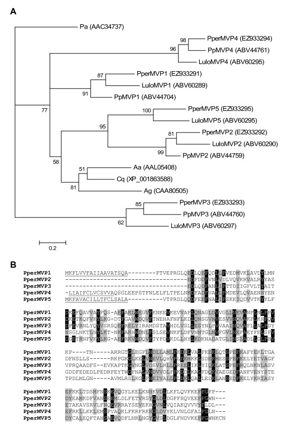
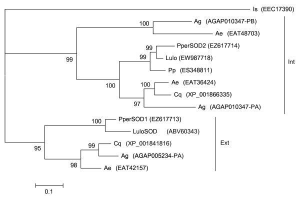
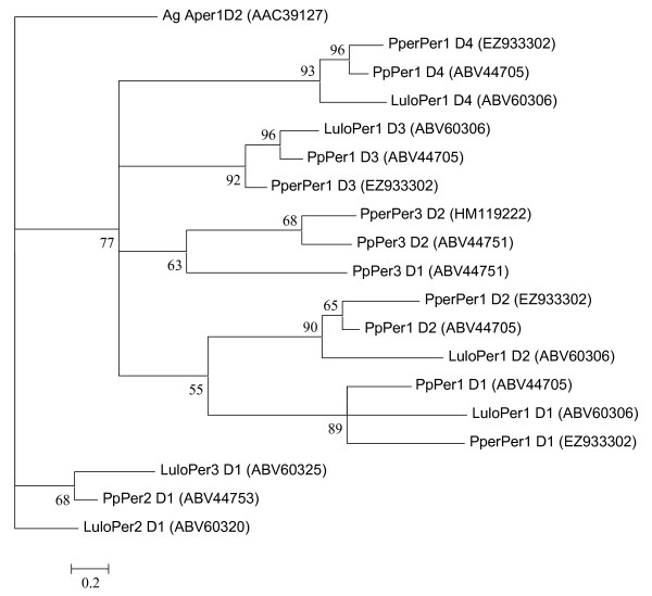
Results: A total of 4111 high quality sequences were obtained from the two libraries and assembled into 370 contigs and 1085 singletons. Molecules with putative roles in blood meal digestion, peritrophic matrix formation, immunity and response to oxidative stress were identified, including proteins that were not previously reported in sand flies. These molecules were evaluated relative to other published sand fly transcripts. Comparative analysis of the two libraries revealed transcripts differentially expressed in response to blood feeding. Molecules up regulated by blood feeding include a putative peritrophin (PperPer1), two chymotrypsin-like proteins (PperChym1 and PperChym2), a putative trypsin (PperTryp3) and four putative microvillar proteins (PperMVP1, 2, 4 and 5). Additionally, several transcripts were more abundant in the sugar fed midgut, such as two putative trypsins (PperTryp1 and PperTryp2), a chymotrypsin (PperChym3) and a microvillar protein (PperMVP3). We performed a detailed temporal expression profile analysis of the putative trypsin transcripts using qPCR and confirmed the expression of blood-induced and blood-repressed trypsins. Trypsin expression was measured in Leishmania infantum-infected and uninfected sand flies, which identified the L. infantum-induced down regulation of PperTryp3 at 24 hours post-blood meal.

Conclusion: This midgut tissue-specific transcriptome provides insight into the molecules expressed in the midgut of P. perniciosus, an important vector of visceral leishmaniasis in the Old World. Through the comparative analysis of the libraries we identified molecules differentially expressed during blood meal digestion. Additionally, this study provides a detailed comparison to transcripts of other sand flies. Moreover, our analysis of putative trypsins demonstrated that L. infantum infection can reduce the transcript abundance of trypsin PperTryp3 in the midgut of P. perniciosus.


Figures

Figure 1
Figure 1
Distribution of clusters from the sugar fed and blood fed libraries in general functional classes. Significant match to the KOG database (E<10E-5) was used as a guideline for grouping the sequences into the functional classes.
Figure 2
Figure 2
Multiple sequence alignment of putative sand fly trypsins. Pper: Phlebotomus perniciosus, Pp: Phlebotomus papatasi, Lulo: Lutzomyia longipalpis. Predicted signal peptides are underlined, the putative activation cleavage site is indicated by (↓), conserved cysteines (C), catalytic H/D/S residues marked by (*) and substrate binding site marked by (#). Accession numbers: PperTryp1 [GenBank:EZ933288], PperTryp2 [GenBank:EZ933289], PperTryp3 [GenBank:EZ933290], Lltryp1 [GenBank:ABM26904], Lltryp2 [GenBank:ABM26905], LuloTryp3 [GenBank:ABV60308], LuloTryp4 [GenBank: ABV60300], PpTryp1 [GenBank:AAM96940], PpTryp2 [GenBank:AAM96941], PpTryp3 [GenBank:AAM96942], PpTryp4 [GenBank:AAM96943].
Figure 3
Figure 3
Influence of blood feeding and L. infantum infection on the dynamics of P. perniciosus trypsins expression. (A) The graph shows PperTryp1, PperTryp2 and PperTryp3 expression as fold over the reference housekeeping gene (PpPerS7 ribosomal protein) before and after the blood feeding (6 hours, 24 hours, 72 hours and 10 days). Each column represents the mean of ten females. S, sugar fed sand flies; B, blood fed sand flies; I, blood fed and L. infantum infected sand flies. The statistically significant difference between the infected and uninfected sand flies is indicated by (*). (B) The graph shows significant difference of PperTryp3 expression in uninfected (B_24) and infected (I_24) sand flies 24 hours after blood feeding; Mann-Whitney U Test (U = 20; Z = 2.268), p = 0.023.
Figure 4
Figure 4
Phylogenetic analysis of putative trypsins from Anopheles stephensi (As), Ochlerotatus taeniorhynchus (Ot), Homo sapiens (Hs), Aedes aegypti (Aa), Anopheles gambiae (Ag), Phlebotomus perniciosus (Pper), Lutzomyia longipalpis (Lulo), Phlebotomus papatasi (Pp) and Drosophila melanogaster (Dm). GenBank accession numbers are given in parentheses and node support is indicated by the bootstrap values.
Figure 5
Figure 5
Phylogenetic analysis of putative chymotrypsin molecules from Litopenaeus vannamei (Lv), Culex quinquefasciatus (Cq), Anopheles gambiae (Ag), Aedes aegypti (Ae), Phlebotomus perniciosus (Pper), Lutzomyia longipalpis (Lulo) and Phlebotomus papatasi (Pp). GenBank accession numbers are given in parentheses and node support indicated by the bootstrap value.
Figure 6
Figure 6
Multiple sequence alignment of putative P. perniciosus chymotrypsins (partial sequences). Conserved cysteines are indicated (C), catalytic H/D/S residues marked by (*) and a serine residue implicated in chymotrypsin substrate specificity marked by (#). Accession numbers: PperChym1 [GenBank:EZ933296], PperChym2 [GenBank:EZ933297], PperChym3 [GenBank:EZ933298], PperChym4 [GenBank:EZ933299], PperChym5 [GenBank:EZ933300].
Figure 7
Figure 7
Phylogenetic analysis of putative carboxypeptidases from Drosophila melanogaster (Dm), Aedes aegypti (Aa), Anopheles gambiae (Ag), Phlebotomus perniciosus (Pper), Lutzomyia longipalpis (Lulo) and Phlebotomus papatasi (Pp). Genbank accession numbers are given in parentheses and node support is indicated by the bootstrap values.
Figure 8
Figure 8
Sequence alignment of putative midgut carboxypeptidases. (A) Comparison of mature Carboxypeptidase A proteins of Aedes aegypti (A. aegypti), Anopheles gambiae (A. gambiae), Phlebotomus perniciosus (Pper), Phlebotomus papatasi (Pp) and Lutzomyia longipalpis (Lulo). N-terminal portion of the peptides are not shown due to PperCpepA 5' mRNA truncation. Conserved cysteines are indicated (C), metal binding residues are marked by (*) and catalytic residues are marked by (#). (B) Comparison of mature Carboxypeptidase B proteins of P. perniciosus (Pper), P. papatasi (Pp) and L. longipalpis (Lulo). Conserved cysteines are indicated (C), metal binding residues are marked by (*), catalytic residues are marked by (#) and a conserved aspartate in the binding pocket of carboxypeptidases B is indicated (D). Accession numbers: PperCpepA [GenBank:EZ966131], A. aegypti [GenBank:AAT36730], A. gambiae [GenBank:AAB96576], LuloCpepA1 [GenBank:ABV60310], LuloCpepA2 [GenBank:ABV60311], PpCpepA [GenBank:ABV44738], PperCpepB [GenBank:EZ966132], PpCpepB [GenBank:ABV44754], LuloCpepB [GenBank:ABV60312].
Figure 9
Figure 9
Phylogenetic analysis and sequence alignment of (putative) astacins. (A) Astacus astacus (Asa), Drosophila melanogaster (Dm), Aedes aegypti (Ae), Anopheles gambiae (Ag), Phlebotomus perniciosus (Pper), Lutzomyia longipalpis (Lulo), Phlebotomus papatasi (Pp) and Culex quinquefasciatus (Cq). Accession numbers are given in parentheses and node support is indicated by the bootstrap values. (B) Drosophila melanogaster (D. melanogaster), Aedes aegypti (A. aegypti), Anopheles gambiae (A. gambiae), Phlebotomus perniciosus (Pper), Lutzomyia longipalpis (Lulo), Phlebotomus papatasi (Pp) and Culex quinquefasciatus (C. quinque). Predicted signal peptide is underlined, conserved cysteines are indicated (C), the metal binding residues marked by (*) and catalytic residues marked by (#). Accession numbers: PperAstacin2 [GenBank:ABV44746], PperAstacin1 [GenBank:EZ966133], LuloAstacin [GenBank:ABV60299], PpAstacin [GenBank:ABV44746], D. melanogaster [GenBank:AAY55427], A. gambiae [GenBank:XP_318553], A. aegypti [GenBank:XP_001648914], C. quinque [GenBank:XP 001844556].
Figure 10
Figure 10
Phylogenetic analysis and sequence alignment of putative microvillar proteins. (A) Periplaneta americana (Pa), Phlebotomus perniciosus (Pper), Phlebotomus papatasi (Pp), Lutzomyia longipalpis (Lulo), Aedes aegypti (Ae), Culex quinquefasciatus (Cq) and Anopheles gambiae (Ag). Accession numbers are given in parentheses. Node support is indicated by the bootstrap values. (B) Phlebotomus perniciosus (Pper). The predicted signal peptides are underlined. Accession numbers: PperMVP1 [GenBank:EZ933291], PperMVP2 [GenBank:EZ933292], PperMVP3 [GenBank:EZ933293], PperMVP4 [GenBank:EZ933294], PperMVP5 [GenBank:EZ933295].
Figure 11
Figure 11
Phylogenetic analysis of superoxide dismutase molecules from Ixodes scapularis (Is), Anopheles gambiae (Ag), Aedes aegypti (Ae), Phlebotomus perniciosus (Pper), Lutzomyia longipalpis (Lulo), Phlebotomus papatasi (Pp) and Culex quinquefasciatus (Cq). GenBank accession numbers are given in parentheses, the clades are labelled with the respective localization based on SignalP prediction (Int: intracellular, Ext: extracellular) and node support is indicated by the bootstrap values.
Figure 12
Figure 12
Phylogenetic analysis of predicted chitin-binding domains of putative peritrophins from Anopheles gambiae (Ag), Phlebotomus perniciosus (Pper), Lutzomyia longipalpis (Lulo) and Phlebotomus papatasi (Pp). GenBank accession numbers are given in parentheses and bootstrap values indicate node support.

References

    1. Killick-Kendrick R. The biology and control of phlebotomine sand flies. Clin Dermatol. 1999;17:279–289. doi: 10.1016/S0738-081X(99)00046-2. - DOI - PubMed
    1. Kamhawi S. Phlebotomine sand flies and Leishmania parasites: friends or foes? Trends Parasitol. 2006;22:439–445. doi: 10.1016/j.pt.2006.06.012. - DOI - PubMed
    1. Peacock CS, Seeger K, Harris D, Murphy L, Ruiz JC, Quail MA, Peters N, Adlem E, Tivey A, Aslett M, Kerhornou A, Ivens A, Fraser A, Rajandream MA, Carver T, Norbertczak H, Chillingworth T, Hance Z, Jagels K, Moule S, Ormond D, Rutter S, Squares R, Whitehead S, Rabbinowitsch E, Arrowsmith C, White B, Thurston S, Bringaud F, Baldauf SL, Faulconbridge A, Jeffares D, Depledge DP, Oyola SO, Hilley JD, Brito LO, Tosi LR, Barrell B, Cruz AK, Mottram JC, Smith DF, Berriman M. Comparative genomic analysis of three Leishmania species that cause diverse human disease. Nat Genet. 2007;39:839–847. doi: 10.1038/ng2053. - DOI - PMC - PubMed
    1. Oliveira F, Jochim RC, Valenzuela JG, Kamhawi S. Sand flies, Leishmania, and transcriptome-borne solutions. Parasitol Int. 2009;58:1–5. doi: 10.1016/j.parint.2008.07.004. - DOI - PMC - PubMed
    1. Ramalho-Ortigao M, Jochim RC, Anderson JM, Lawyer PG, Pham VM, Kamhawi S, Valenzuela JG. Exploring the midgut transcriptome of Phlebotomus papatasi: comparative analysis of expression profiles of sugarfed, blood-fed and Leishmania major-infected sandflies. BMC Genomics. 2007. p. 8. - PMC - PubMed

Publication types

MeSH terms