. 2011 May 10:6:24.
doi: 10.1186/1749-8104-6-24.

Coordinated trafficking of synaptic vesicle and active zone proteins prior to synapse formation

Affiliations

Coordinated trafficking of synaptic vesicle and active zone proteins prior to synapse formation

Luke A D Bury et al. Neural Dev. .

Abstract

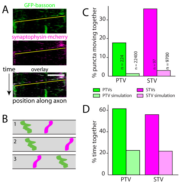
Background: The proteins required for synaptic transmission are rapidly assembled at nascent synapses, but the mechanisms through which these proteins are delivered to developing presynaptic terminals are not understood. Prior to synapse formation, active zone proteins and synaptic vesicle proteins are transported along axons in distinct organelles referred to as piccolo-bassoon transport vesicles (PTVs) and synaptic vesicle protein transport vesicles (STVs), respectively. Although both PTVs and STVs are recruited to the same site in the axon, often within minutes of axo-dendritic contact, it is not known whether or how PTV and STV trafficking is coordinated before synapse formation.

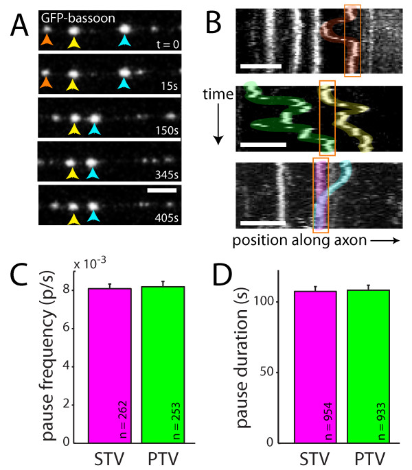
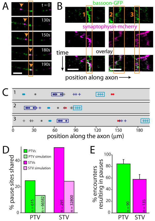
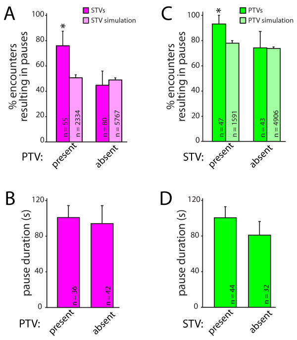
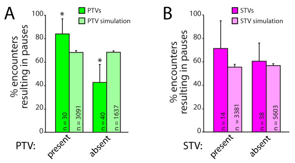
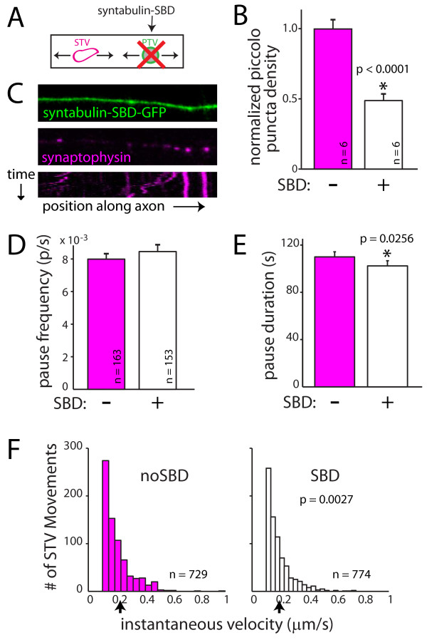
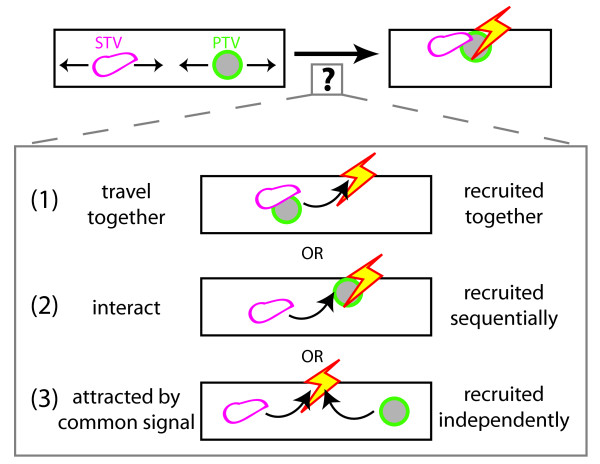
Results: Here, using time-lapse confocal imaging of the dynamics of PTVs and STVs in the same axon, we show that vesicle trafficking is coordinated through at least two mechanisms. First, a significant proportion of STVs and PTVs are transported together before forming a stable terminal. Second, individual PTVs and STVs share pause sites within the axon. Importantly, for both STVs and PTVs, encountering the other type of vesicle increases their propensity to pause. To determine if PTV-STV interactions are important for pausing, PTV density was reduced in axons by expression of a dominant negative construct corresponding to the syntaxin binding domain of syntabulin, which links PTVs with their KIF5B motor. This reduction in PTVs had a minimal effect on STV pausing and movement, suggesting that an interaction between STVs and PTVs is not responsible for enhancing STV pausing.

Conclusions: Our results indicate that trafficking of STVs and PTVs is coordinated even prior to synapse development. This novel coordination of transport and pausing might provide mechanisms through which all of the components of a presynaptic terminal can be rapidly accumulated at sites of synapse formation.


Figures

Figure 1
Figure 1
STVs and PTVs move together. (A) Kymographs of an axon segment showing the movements of PTVs (GFP-bassoon; green) and STVs (synaptophysin-mcherry; magenta) in the same axon. Yellow lines underline the moving STVs and PTVs. Bottom panel: overlay of STV and PTV fluorescence. On the ordinate axis, one pixel corresponds to 10 s. On the abscissa, the scale bar corresponds to 10 μm. (B) Simulations of model axons were performed by randomizing initial positions of vesicles while maintaining movement and pausing characteristics of the original imaged vesicles. Diagrams illustrate kymographs of three simulations of model axons. Movements of two vesicles are shown in each model axon. (C) Plot showing the percentage of PTVs that move with STVs (green) and STVs that move with PTVs (magenta). PTV and STV simulations (light green and light magenta, respectively) correspond to the predicted values from the simulations. (D) Quantification of the percentage of time that PTVs and STVs spent together. Both the percentage of vesicles that moved together and the time spent together were substantially larger in the data than is predicted by chance (simulations).
Figure 2
Figure 2
PTV pausing is qualitatively and quantitatively similar to STV pausing. (A) Time-lapse images of PTVs labeled with GFP-bassoon. Individual PTVs are tracked with red, yellow or blue arrows. Most PTVs paused, and pauses were of varied durations. Frames were collected at the indicated times. Scale bars: 5 μm. (B) Kymographs demonstrating the movements of PTVs in three axons. Individual PTVs paused repeatedly at the same site (top panel), and multiple PTVs paused at the same site (middle and bottom panels). Pausing of multiple PTVs at a given site occurred both sequentially (middle panel) and simultaneously (bottom panel). Individual vesicles are highlighted in different colors for visualization. Pause sites are outlined in orange. On the ordinate axis, one pixel corresponds to 10 s. (C,D) STVs (magenta) and PTVs (green) paused at similar frequencies (C) and for similar mean durations (D). Data represent the mean + standard error.
Figure 3
Figure 3
STVs and PTVs pause at the same sites. (A) Time-lapse images of a segment of axon expressing both synaptophysin-mcherry (magenta) and GFP-bassoon (green). In each panel, fluorescence signals from the two channels are overlaid, and co-localization is indicated by white. The orange box outlines a site where a PTV is paused. The arrow tracks a STV that then pauses at that site. Scale bars: 5 μm. (B) Kymographs showing two examples of STVs and PTVs that pause at the same sites. On the ordinate axis, one pixel corresponds to 10 s. Bottom panel: overlay of STV and PTV fluorescence. Orange boxes outline three shared pause sites. (C) Simulations of model axons were performed by randomizing initial positions of vesicles while maintaining movement and pausing characteristics observed for the original experimental vesicles. Diagrams show the superimposed locations of all pause sites for individual STVs and PTVs in model axons. Three simulations of the same experimental data are shown. STV pause sites are indicated with a dot while PTV pause sites are indicated with a plus sign. Each color represents an individual vesicle. Cyan and magenta boxes outline the pause sites of the same PTV and STV, respectively, in each model axon. For each axon imaged, 100 such simulations were performed, allowing estimation of the degree of co-transport and co-pausing expected from chance alone. (D) Plot illustrating the percentage of PTV (green) and STV (magenta) pause sites that are shared with STVs and PTVs, respectively. The fraction of shared sites is much higher than predicted by chance (via simulations, light green and light magenta). (E) A large majority of PTVs that encountered STV pause sites paused at those sites. Similarly, most STVs that encountered PTV pause sites then paused at those sites. Error bars display the 95% confidence interval.
Figure 4
Figure 4
STVs and PTVs preferentially pause at the same sites at the same time. (A) STVs (magenta) are significantly more likely to stop at a PTV pause site when PTVs are at the site. This preference cannot be accounted for by chance since no dependence on the presence of PTVs was seen in simulations (light magenta). The data presented correspond to the mean + 95% confidence intervals; *95% confidence intervals do not overlap. (B) STVs pause for similar lengths of time regardless of whether a PTV is present at the pause site. Data are presented as mean + standard error. (C) PTVs (green) are more likely to pause at a site when an STV is present. The same dependence was not observed in simulations (light green). (D) PTVs pause for similar lengths of time, regardless of whether STVs are present at the pause sites. Data are the mean + standard error.
Figure 5
Figure 5
Multiple PTVs are attracted to the same sites. (A) PTVs are more likely to pause when other PTVs are present (green). Spatially randomized simulations are shown in light green and cannot account for the tendency of PTVs to pause simultaneously with other PTVs. (B) STVs are not more likely to pause when other STVs are already present (magenta). Simulations are shown for comparison (light magenta). Error bars represent 95% confidence intervals; *95% confidence intervals do not overlap.
Figure 6
Figure 6
A direct interaction between STVs and PTVs cannot account for the attraction of STVs to pause sites that contain PTVs. (A) PTV transport was inhibited using dominant-negative syntabulin (syntaxin binding domain, SBD) fused to GFP. (B) Expression of syntabulin-SBD-GFP decreases the density of PTV puncta in axons when compared to axons expressing GFP alone. PTVs were identified by immunofluorescent labeling for endogenous piccolo. (C) Images and kymograph (bottom) showing that STVs (magenta) move and pause in SBD-expressing axons (green). (D) The frequency of STV pausing was unchanged in the presence of SBD-GFP. (E) STV pause durations are shorter when PTV localization in the axon is disrupted. The change in pausing upon expression of SBD-GFP is not sufficient to account for the coordinated pausing of STVs and PTVs. In B, D and E, error bars correspond to the standard error. *, difference is significant. P-values are from Wilcoxon rank-sum test and are indicated in the figure. (F) The instantaneous velocities of STVs are increased in SBD-expressing axons. Black arrow, mean instantaneous velocity.
Figure 7
Figure 7
Models for coordination of recruitment of STVs and PTVs to the same place at the same time. STVs and PTVs could be simultaneously attracted to a given site by (1) attraction of STVs and PTVs that are co-transported; (2) sequential recruitment via a physical interaction between STVs and PTVs; and (3) simultaneous but independent recruitment in response to a common signal. Our data are most consistent with the third model.

References

    1. Bury LA, Sabo SL. How it's made: the synapse. Mol Interv. 2010;10:282–292. doi: 10.1124/mi.10.5.5. - DOI - PubMed
    1. McAllister AK. Dynamic aspects of CNS synapse formation. Annu Rev Neurosci. 2007;30:425–450. doi: 10.1146/annurev.neuro.29.051605.112830. - DOI - PMC - PubMed
    1. Friedman HV, Bresler T, Garner CC, Ziv NE. Assembly of new individual excitatory synapses: time course and temporal order of synaptic molecule recruitment. Neuron. 2000;27:57–69. doi: 10.1016/S0896-6273(00)00009-X. - DOI - PubMed
    1. Sabo SL, Gomes RA, McAllister AK. Formation of presynaptic terminals at predefined sites along axons. J Neurosci. 2006;26:10813–10825. doi: 10.1523/JNEUROSCI.2052-06.2006. - DOI - PMC - PubMed
    1. Washbourne P, Bennett JE, McAllister AK. Rapid recruitment of NMDA receptor transport packets to nascent synapses. Nat Neurosci. 2002;5:751–759. - PubMed

Publication types

Substances

LinkOut - more resources