. 2011 May 11:12:40.
doi: 10.1186/1471-2202-12-40.

The role of shear stress in Blood-Brain Barrier endothelial physiology

Affiliations

The role of shear stress in Blood-Brain Barrier endothelial physiology

Luca Cucullo et al. BMC Neurosci. .

Abstract

Background: One of the most important and often neglected physiological stimuli contributing to the differentiation of vascular endothelial cells (ECs) into a blood-brain barrier (BBB) phenotype is shear stress (SS). With the use of a well established humanized dynamic in vitro BBB model and cDNA microarrays, we have profiled the effect of SS in the induction/suppression of ECs genes and related functions.

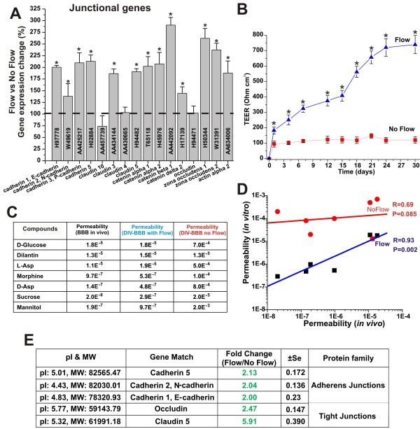
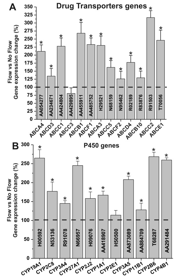
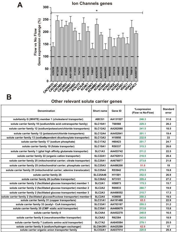
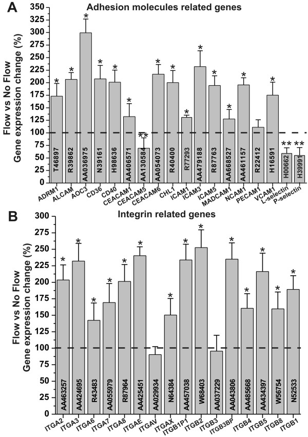
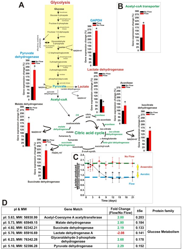
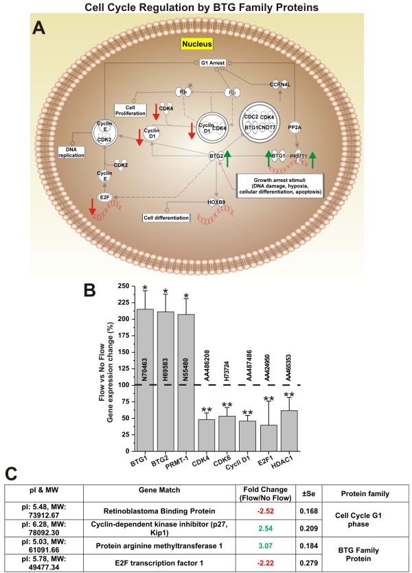
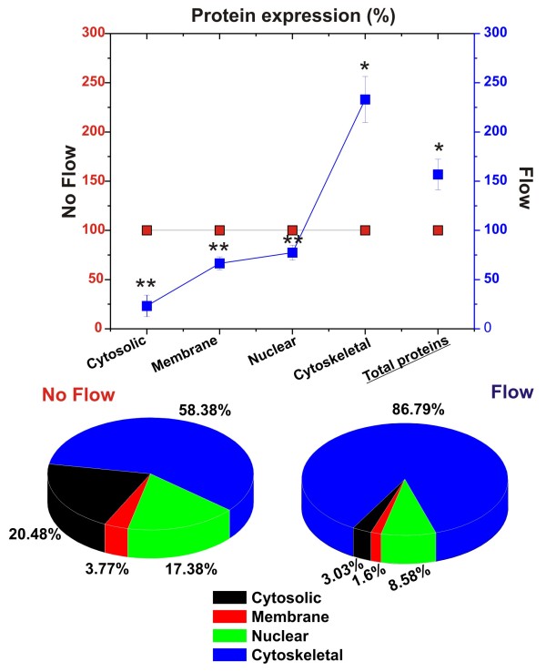
Results: Specifically, we found a significant upregulation of tight and adherens junctions proteins and genes. Trans-endothelial electrical resistance (TEER) and permeability measurements to know substances have shown that SS promoted the formation of a tight and highly selective BBB. SS also increased the RNA level of multidrug resistance transporters, ion channels, and several p450 enzymes. The RNA level of a number of specialized carrier-mediated transport systems (e.g., glucose, monocarboxylic acid, etc.) was also upregulated.RNA levels of modulatory enzymes of the glycolytic pathway (e.g., lactate dehydrogenase) were downregulated by SS while those involved in the Krebs cycle (e.g., lactate and other dehydrogenases) were upregulated. Measurements of glucose consumption versus lactate production showed that SS negatively modulated the glycolytic bioenergetic pathways of glucose metabolism in favor of the more efficient aerobic respiration. BBB ECs are responsive to inflammatory stimuli. Our data showed that SS increased the RNA levels of integrins and vascular adhesion molecules. SS also inhibited endothelial cell cycle via regulation of BTG family proteins encoding genes. This was paralleled by significant increase in the cytoskeletal protein content while that of membrane, cytosol, and nuclear sub-cellular fractions decreased. Furthermore, analysis of 2D gel electrophoresis (which allows identifying a large number of proteins per sample) of EC proteins extracted from membrane sub-cellular endothelial fractions showed that SS increased the expression levels of tight junction proteins. In addition, regulatory enzymes of the Krebb's cycle (aerobic glucose metabolism) were also upregulated. Furthermore, the expression pattern of key protein regulators of the cell cycle and parallel gene array data supported a cell proliferation inhibitory role for SS.

Conclusions: Genomic and proteomic analyses are currently used to examine BBB function in healthy and diseased brain and characterize this dynamic interface. In this study we showed that SS plays a key role in promoting the differentiation of vascular endothelial cells into a truly BBB phenotype. SS affected multiple aspect of the endothelial physiology spanning from tight junctions formation to cell division as well as the expression of multidrug resistance transporters. BBB dysfunction has been observed in many neurological diseases, but the causes are generally unknown. Our study provides essential insights to understand the role played by SS in the BBB formation and maintenance.


Figures

Figure 1
Figure 1
Laminar shear stress promotes tight junction formation. Panel A: Comparison of the level of TJ and adherens junction RNA in EC grown under dynamic (flow) versus static condition. The tightness of the vascular endothelial bed developed under flow culture condition was much more stringent than that grown under in the absence of luminal flow. This was demonstrated by the TEER (Panel B) and permeability measurement to known substances (Panel C). Note also that the BBB established under flow was capable of discriminating the passage of substances accordingly to their permeability order more efficiently that the BBB established under static condition (Panel D). Gene array findings were also supported by comparative analysis of TJ and adherens junction proteins identified and quantified (comparative analysis of the expression levels) on 2D gels of protein extracted from EC membrane sub-cellular fractions (flow and no-flow conditions; Panel E). Note that "*" refers to a statistically significant changes in BBB tightness and EC junctional genes expression caused by the exposure to flow (n = 4; p < 0.05).
Figure 2
Figure 2
Shear stress enables vascular-dependent shielding of brain from unwanted and potentially harmful substances. Shear stress increases the RNA levels of gene encoding for multidrug resistance proteins (panel A) and cytochrome P450 enzymes (panel B). Note that "*" refers to a statistically significant (n = 4; p < 0.05) increase of the genes transcription in EC exposed to flow versus those grown under static conditions.
Figure 3
Figure 3
Shear stress enhances the RNA levels of ion channel genes and other relevant transporters. Note that "*" refers to a statistically significant (n = 4; p < 0.05) increase of the genes transcription in EC exposed to flow versus those grown under static conditions. Note also that the data in panel B are reported as % of the RNA level measured in EC grown under static condition ± SEM.
Figure 4
Figure 4
Shear stress enables endothelial-leukocyte interaction. Shear stress increased the transcription level of relevant adhesion molecules (Panel A) and integrins (Panel B) thus facilitating the cross-talk between the vascular endothelium and the host immune system at the BBB level. Note that "*" and "**" refers to a statistically significant (n = 4; p < 0.05) changes (increase and decrease respectively) of adhesion molecules and integrins genes transcription in EC exposed to flow versus those grown under static conditions.
Figure 5
Figure 5
Shear stress modulates the endothelial bioenergetic pathway. Panel A and B Enzimatic pathways of glycolysis and Krebb's cycle. Note the change in the expression of genes encoding for the key regulator enzymes of glucose metabolism and how the exposure to flow greatly favoured the more efficient aerobic pathway. The gene array data were confirmed by measurements of glucose consumption versus lactate production in DIV-BBB model developed with and without l endothelial exposure to intraluminal flow (Panel C). Comparative analysis of the expression level of key enzymes regulating the glycolitic and the aerobic (Krebb's cycle) pathways strongly supported the gene array data (Panel D). Note that the lactate production/glucose consumption ratio measured in the flow-exposed in vitro BBB modules was ≈ 1. Complete anaerobic metabolism would produce 2 lactates/glucose (ratio = 2) thus, indicating that at least 50% of the glucose consumed underwent aerobic metabolism.
Figure 6
Figure 6
Shear stress inhibits cell proliferation. Panel A: schematic representation of the cell cycle regulation by BTG family proteins. Functional correlation between gene expression and cell cycle was determined using Pathway analysis software. Note how shear stress increased the RNA levels of genes encoding the cell cycle inhibitory effectors BTG1, BTG2 and PMRT1 (Panel B). This finding was also supported by the comparative analysis (flow vs. no-flow) of the expression level of cell cycle modulatory proteins (Panel C). Note that genes and protein related to positive modulators of the cell cycle progression were significantly downregulated. Note also that "*" and "**" refers to a statistically significant (n = 4; p < 0.05) changes (increase and decrease respectively).
Figure 7
Figure 7
Shear stress increases cytoskeleton protein expression while decreasing that of cytosol, nucleus and membrane. Note the drastic changes related to cytoskeletal protein expression. In EC grown under flow condition cytoskeletal proteins represent account for majority of the total protein content. Note also that the exposure to flow significantly decreased the overall expression of nuclear, cytosolic and membrane proteins. This is consistent with endothelial morphologic adaptation to flow and with a reduced cell proliferation. Note that "*" and "**" refers to a statistically significant (n = 4; P < 0.05) changes (increase and decrease respectively) in the protein content.

References

    1. Emmi A, Wenzel HJ, Schwartzkroin PA, Taglialatela M, Castaldo P, Bianchi L. et al. Do glia have heart? Expression and functional role for ether-a-go-go currents in hippocampal astrocytes. J Neurosci. 2000;20:3915–3925. - PMC - PubMed
    1. Cornford EM, Hyman S. Localization of brain endothelial luminal and abluminal transporters with immunogold electron microscopy. NeuroRx. 2005;2:27–43. doi: 10.1602/neurorx.2.1.27. - DOI - PMC - PubMed
    1. Dauchy S, Dutheil F, Weaver RJ, Chassoux F, Daumas-Duport C, Couraud PO. et al. ABC transporters, cytochromes P450 and their main transcription factors: expression at the human blood-brain barrier. J Neurochem. 2008;107:1518–1528. doi: 10.1111/j.1471-4159.2008.05720.x. - DOI - PubMed
    1. Cornford EM, Varesi JB, Hyman S, Damian RT, Raleigh MJ. Mitochondrial content of choroid plexus epithelium. Exp Brain Res. 1997;116:399–405. doi: 10.1007/PL00005768. - DOI - PubMed
    1. Steiner O, Coisne C, Cecchelli R, Boscacci R, Deutsch U, Engelhardt B. et al. Differential roles for endothelial ICAM-1, ICAM-2, and VCAM-1 in shear-resistant T cell arrest, polarization, and directed crawling on blood-brain barrier endothelium. J Immunol. 2010;185:4846–4855. doi: 10.4049/jimmunol.0903732. - DOI - PubMed

Publication types

LinkOut - more resources