. 2011;15(3):R124.
doi: 10.1186/cc10230. Epub 2011 May 13.

Injurious mechanical ventilation affects neuronal activation in ventilated rats

Affiliations

Injurious mechanical ventilation affects neuronal activation in ventilated rats

María Elisa Quilez et al. Crit Care. 2011.

Erratum in

Abstract

Introduction: Survivors of critical illness often have significant long-term brain dysfunction, and routine clinical procedures like mechanical ventilation (MV) may affect long-term brain outcome. We aimed to investigate the effect of the increase of tidal volume (Vt) on brain activation in a rat model.

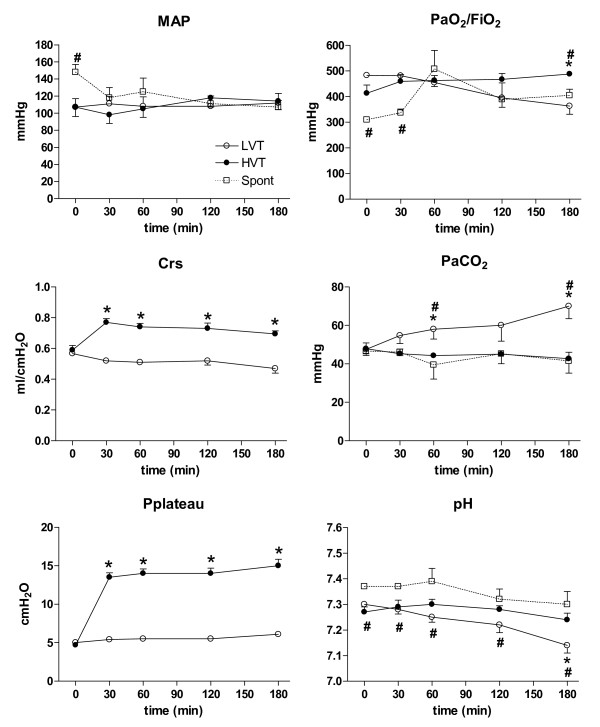
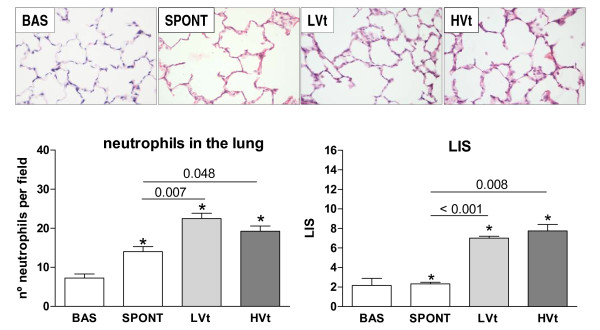
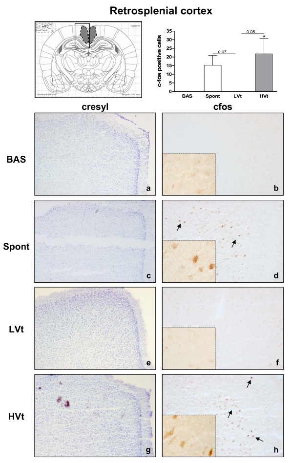
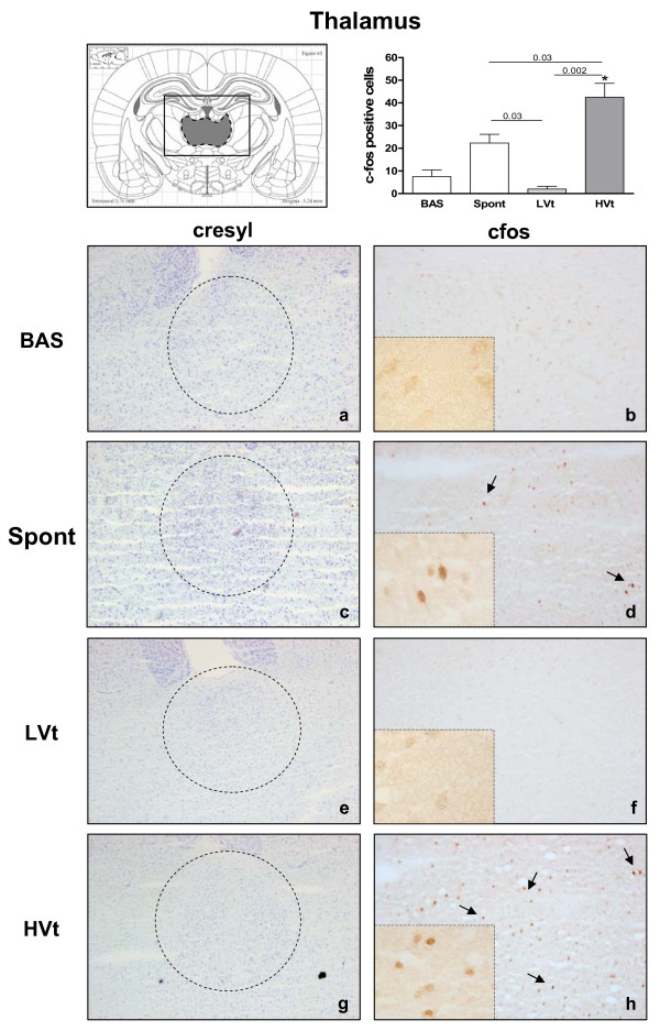
Methods: Male Sprague Dawley rats were randomized to three groups: 1) Basal: anesthetized unventilated animals, 2) low Vt (LVt): MV for three hours with Vt 8 ml/kg and zero positive end-expiratory pressure (ZEEP), and 3) high Vt (HVt) MV for three hours with Vt 30 ml/kg and ZEEP. We measured lung mechanics, mean arterial pressure (MAP), arterial blood gases, and plasma and lung levels of cytokines. We used immunohistochemistry to examine c-fos as a marker of neuronal activation. An additional group of spontaneously breathing rats was added to discriminate the effect of surgical procedure and anesthesia in the brain.

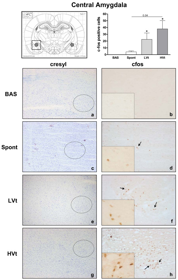
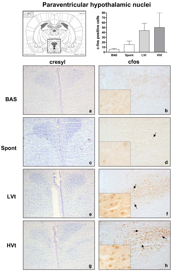
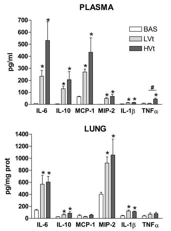
Results: After three hours on LVt, PaO2 decreased and PaCO2 increased significantly. MAP and compliance remained stable in MV groups. Systemic and pulmonary inflammation was higher in MV rats than in unventilated rats. Plasma TNFα was significantly higher in HVt than in LVt. Immunopositive cells to c-fos in the retrosplenial cortex and thalamus increased significantly in HVt rats but not in LVt or unventilated rats.

Conclusions: MV promoted brain activation. The intensity of the response was higher in HVt animals, suggesting an iatrogenic effect of MV on the brain. These findings suggest that this novel cross-talking mechanism between the lung and the brain should be explored in patients undergoing MV.


Figures

Figure 1
Figure 1
Hemodynamic and respiratory characteristics of rats during the three-hour period. No differences between groups were observed at baseline. MAP remained stable in both groups. Pplateau and Crs increased significantly during HVt ventilation but remained stable during the three-hour period. There were no differences between LVt and HVt in Pa/FiO2. pH in animals spontaneously breathing was slightly higher than in animals receiving MV. PCO2 increased only in LVt animals. Data are presented as mean ± SE. *: P < 0.05 versus the HVt group, and #: P < 0.05 vs Spont group. N = 8 animals per group. Abbreviations: MAP, mean arterial pressure; BAS, basal; LVt, low tidal volume; HVt, high tidal volume; Spont, spontaneous breathing; Pplateau, plateau pressure; Crs, static compliance of the respiratory system.
Figure 2
Figure 2
Representative images of lungs in each group after H-E staining and LIS. The percent of lung neutrophil content and LIS increased with MV but was similar in animals receiving LVt and HVt. Results are represented as mean ± SE. *P < 0.05 versus the unventilated basal group. N = 8 animals per group. Abbreviations: BAS, basal; LVt, low tidal volume; HVt, high tidal volume; Spont, spontaneous breathing; LIS, Lung injury score.
Figure 3
Figure 3
Brain activation evidenced by c-fos immunotreactivity in the retrosplenial cortex. On the top (left): Coronal section diagram encompassing the area of interest. On the bottom: Representative images of RS from each experimental group after cresyl violet staining (left, a,c,e,g, 40X) and c-fos immunohistochemistry (right 100X and 400X). Brown dots represent c-fos staining. Black arrows indicate some examples of c-fos positive cells. HVt (h) and spontaneous breathing (d) increased the number of c-fos-positive neurons in the RS; lower levels of c-fos immunoreactive cells were found in unventilated (b) and LVt (f) animals. Data are presented as mean ± SE. *P < 0.05 respect to unventilated basal animals. Abbreviations: BAS, basal; LVt, low tidal volume; HVt, high tidal volume; Spont, spontaneous breathing; RS, retrosplenial cortex.
Figure 4
Figure 4
Brain activation evidenced by c-fos immunotreactivity in thalamus. On the top (left): Coronal section diagram encompassing the area of interest. On the bottom: Representative images of thalamus from each experimental group after cresyl violet staining (left, a,c,e,g, 40X) and c-fos immunohistochemistry (right 100X and 400X). Brown dots represent c-fos staining. Black arrows indicate some examples of c-fos positive cells. HVt (h) and spontaneous breathing (d) increased the number of c-fos-positive neurons in the thalamus. Data are presented as mean ± SE. *P < 0.05 respect to unventilated basal animals. Abbreviations: BAS, basal; LVt, low tidal volume; HVt, high tidal volume; Spont, spontaneous breathing.
Figure 5
Figure 5
Brain activation evidenced by c-fos immunotreactivity in central amygdala. On the top (left): Coronal section diagram encompassing the area of interest. On the bottom: Representative images of the central amygdala from each experimental group after cresyl violet staining (left,a, c,e,g, 40X) and c-fos immunohistochemistry (right, 100X and 400X). Brown dots represent c-fos staining. Black arrows indicate some examples of c-fos positive cells. MV significantly increased c-fos immunoreactive cells in the CeA independently of the Vt level (f, h ). Few c-fos positive cells were found in CeA of spontaneous breathing (d) and basal (b) animals. Data are presented as mean ± SE. *P < 0.05 respect to unventilated basal animals. Abbreviations: No MV, unventilated animals; LVt, low tidal volume; HVt, high tidal volume; Spont, spontaneous breathing; CeA, central amygdala.
Figure 6
Figure 6
Brain activation evidenced by c-fos immunotreactivity in Paraventricular hypothalamic nuclei. On the top (left): Coronal section diagram encompassing the area of interest. On the bottom: Representative images of Paraventricular hypothalamic nuclei from each experimental group after cresyl violet staining (left, a,c,e,g, 40X) and c-fos immunohistochemistry (right 100X and 400X). Brown dots represent c-fos staining. Black arrows indicate some examples of c-fos positive cells c-fos expression in the PVN tended to increase with MV (f, h), but this increase did not reach significance compared with basal (b) or spontaneous breathing rats (d). Data are presented as mean ± SE. *P < 0.05 respect to unventilated basal animals. Abbreviations: BAS, basal; LVt, low tidal volume; HVt, high tidal volume; Spont, spontaneous breathing; PVN, Paraventricular hypothalamic nuclei.
Figure 7
Figure 7
Plasma and lung levels of proteins involved in the inflammatory cascade. Mechanical ventilation triggered lung and systemic inflammatory responses. Compared to LVt, HVt promoted an increase in inflammatory markers mainly mediated by TNFα at the plasma leve. Data are presented as mean ± SE. *P < 0.05 respect to unventilated basal animals, # P < 0.05 vs LVt. n = 8 animals per group. Abbreviations: BAS, basal; LVt, low tidal volume; HVt, high tidal volume; Spont, spontaneous breathing; IL, interleukin, TNF, tumor necrosis factor; MCP, monocyte chemotactic protein; MIP, macrophage-inflammatory protein.

References

    1. Rubenfeld GD, Caldwell E, Peabody E, Weaver J, Martin DP, Neff M, Stern EJ, Hudson LD. Incidence and outcomes of acute lung injury. N Engl J Med. 2005;353:1685–1693. doi: 10.1056/NEJMoa050333. - DOI - PubMed
    1. Hopkins RO, Jackson JC. Long-term neurocognitive function after critical illness. Chest. 2006;130:869–878. doi: 10.1378/chest.130.3.869. - DOI - PubMed
    1. Jackson JC, Girard TD, Gordon SM, Thompson JL, Shintani AK, Thomason JW, Pun BT, Canonico AE, Dunn JG, Bernard GR, Dittus RS, Ely EW. Long-term cognitive and psychological outcomes in the Awakening and Breathing Controlled trial. Am J Respir Crit Care Med. 2010;182:183–191. doi: 10.1164/rccm.200903-0442OC. - DOI - PMC - PubMed
    1. Wolthuis EK, Vlaar AP, Choi G, Roelofs JJ, Juffermans NP, Schultz MJ. Mechanical ventilation using non-injurious ventilation settings causes lung injury in the absence of pre-existing lung injury in healthy mice. Crit Care. 2009;13:R1. doi: 10.1186/cc7688. - DOI - PMC - PubMed
    1. Vaneker M, Joosten LA, Heunks LM, Snijdelaar DG, Halbertsma FJ, van Egmond J, Netea MG, van der Hoeven JG, Scheffer GJ. Low-tidal-volume mechanical ventilation induces a toll-like receptor 4-dependent inflammatory response in healthy mice. Anesthesiology. 2008;109:465–472. doi: 10.1097/ALN.0b013e318182aef1. - DOI - PubMed

Publication types

LinkOut - more resources