

Galanin-immunoreactivity identifies a distinct population of inhibitory interneurons in laminae I-III of the rat spinal cord
- PMID: 21569622
- PMCID: PMC3118366
- DOI: 10.1186/1744-8069-7-36
Galanin-immunoreactivity identifies a distinct population of inhibitory interneurons in laminae I-III of the rat spinal cord
Abstract
Background: Inhibitory interneurons constitute 30-40% of neurons in laminae I-III and have an important anti-nociceptive role. However, because of the difficulty in classifying them we know little about their organisation. Previous studies have identified 3 non-overlapping groups of inhibitory interneuron, which contain neuropeptide Y (NPY), neuronal nitric oxide synthase (nNOS) or parvalbumin, and have shown that these differ in postsynaptic targets. Some inhibitory interneurons contain galanin and the first aim of this study was to determine whether these form a different population from those containing NPY, nNOS or parvalbumin. We also estimated the proportion of neurons and GABAergic axons that contain galanin in laminae I-III.
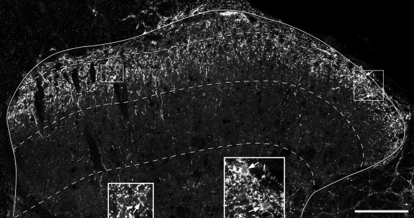
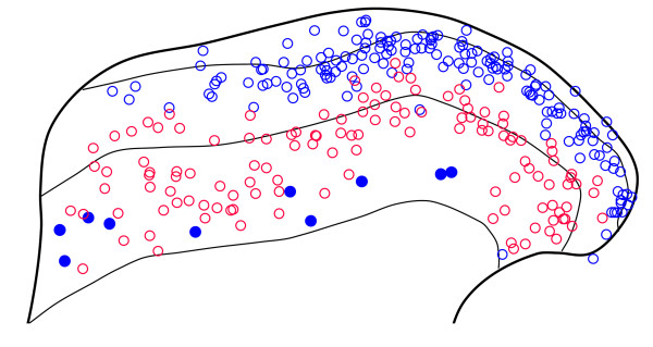
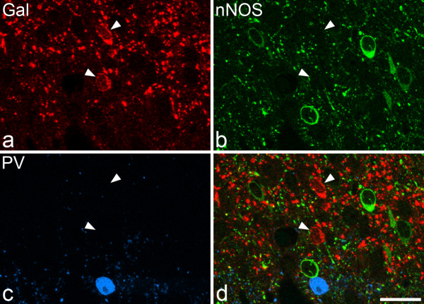
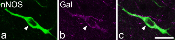
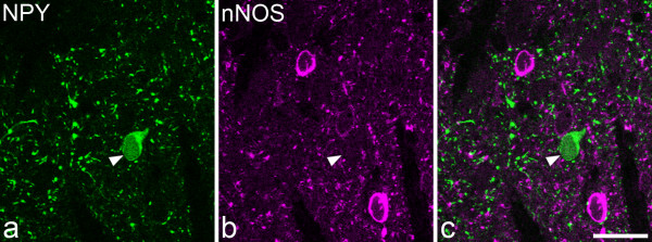
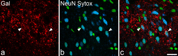
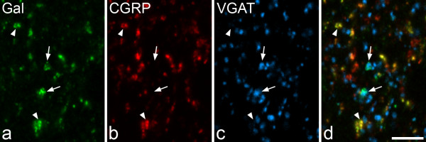
Results: Galanin cells were concentrated in laminae I-IIo, with few in laminae IIi-III. Galanin showed minimal co-localisation with NPY, nNOS or parvalbumin in laminae I-II, but most galanin-containing cells in lamina III were nNOS-positive. Galanin cells constituted ~7%, 3% and 2% of all neurons in laminae I, II and III, and we estimate that this corresponds to 26%, 10% and 5% of the GABAergic neurons in these laminae. However, galanin was only found in ~6% of GABAergic boutons in laminae I-IIo, and ~1% of those in laminae IIi-III.
Conclusions: These results show that galanin, NPY, nNOS and parvalbumin can be used to define four distinct neurochemical populations of inhibitory interneurons. Together with results of a recent study, they suggest that the galanin and NPY populations account for around half of the inhibitory interneurons in lamina I and a quarter of those in lamina II.
Figures

Similar articles
-
Quantitative study of NPY-expressing GABAergic neurons and axons in rat spinal dorsal horn.J Comp Neurol. 2011 Apr 15;519(6):1007-23. doi: 10.1002/cne.22570. J Comp Neurol. 2011. PMID: 21344400 Free PMC article.
-
Functional differences between neurochemically defined populations of inhibitory interneurons in the rat spinal dorsal horn.Pain. 2013 Dec;154(12):2606-2615. doi: 10.1016/j.pain.2013.05.001. Epub 2013 May 7. Pain. 2013. PMID: 23707280 Free PMC article.
-
Dynorphin is expressed primarily by GABAergic neurons that contain galanin in the rat dorsal horn.Mol Pain. 2011 Sep 29;7:76. doi: 10.1186/1744-8069-7-76. Mol Pain. 2011. PMID: 21958458 Free PMC article.
-
The neuropeptide tyrosine Y1R is expressed in interneurons and projection neurons in the dorsal horn and area X of the rat spinal cord.Neuroscience. 2006;138(4):1361-76. doi: 10.1016/j.neuroscience.2005.11.069. Epub 2006 Jan 31. Neuroscience. 2006. PMID: 16448775
-
A quantitative study of neuronal nitric oxide synthase expression in laminae I-III of the rat spinal dorsal horn.Neuroscience. 2011 Sep 29;192(6-2):708-20. doi: 10.1016/j.neuroscience.2011.07.011. Epub 2011 Jul 14. Neuroscience. 2011. PMID: 21763759 Free PMC article.
Cited by
-
HCN4 subunit expression in fast-spiking interneurons of the rat spinal cord and hippocampus.Neuroscience. 2013 May 1;237:7-18. doi: 10.1016/j.neuroscience.2013.01.028. Epub 2013 Jan 26. Neuroscience. 2013. PMID: 23357121 Free PMC article.
-
Neural processing of itch.Neuroscience. 2013 Oct 10;250:697-714. doi: 10.1016/j.neuroscience.2013.07.035. Epub 2013 Jul 24. Neuroscience. 2013. PMID: 23891755 Free PMC article. Review.
-
Dual-transmitter systems regulating arousal, attention, learning and memory.Neurosci Biobehav Rev. 2018 Feb;85:21-33. doi: 10.1016/j.neubiorev.2017.07.009. Epub 2017 Jul 27. Neurosci Biobehav Rev. 2018. PMID: 28757457 Free PMC article. Review.
-
Neurochemical atlas of the rabbit spinal cord.Brain Struct Funct. 2024 Nov;229(8):2011-2027. doi: 10.1007/s00429-024-02842-z. Epub 2024 Aug 8. Brain Struct Funct. 2024. PMID: 39115602
-
Genome-wide expression analysis of Ptf1a- and Ascl1-deficient mice reveals new markers for distinct dorsal horn interneuron populations contributing to nociceptive reflex plasticity.J Neurosci. 2013 Apr 24;33(17):7299-307. doi: 10.1523/JNEUROSCI.0491-13.2013. J Neurosci. 2013. PMID: 23616538 Free PMC article.
References
-
- Polgár E, Hughes DI, Riddell JS, Maxwell DJ, Puskar Z, Todd AJ. Selective loss of spinal GABAergic or glycinergic neurons is not necessary for development of thermal hyperalgesia in the chronic constriction injury model of neuropathic pain. Pain. 2003;104:229–239. doi: 10.1016/S0304-3959(03)00011-3. - DOI - PubMed
-
- Sivilotti L, Woolf CJ. The contribution of GABAA and glycine receptors to central sensitization: disinhibition and touch-evoked allodynia in the spinal cord. J Neurophysiol. 1994;72:169–179. - PubMed
Publication types
MeSH terms
Substances
Grants and funding
LinkOut - more resources
Full Text Sources
Miscellaneous
