. 2011 Aug 1;57(3):817-24.
doi: 10.1016/j.neuroimage.2011.04.063. Epub 2011 May 6.

Serial monitoring of endogenous neuroblast migration by cellular MRI

Affiliations

Serial monitoring of endogenous neuroblast migration by cellular MRI

Dorit Granot et al. Neuroimage. .

Abstract

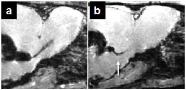
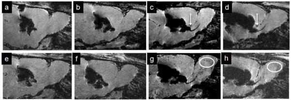
Endogenous neural progenitor cell migration in vivo can be monitored using MRI-based cell tracking. The current protocol is that micron sized iron oxide particles (MPIOs) are injected into the lateral ventricle proximal to the neural stem cell niche in the brain. MPIOs are endocytosed and incorporated into the neural progenitor cell population, making them visible by gradient echo MRI. Here this new method is extended to serially quantify cell migration. Initially, in vivo cell labeling methodologies were optimized, as high susceptibility effects from the MPIOs generate substantial signal loss around the injection site, masking early migratory events. Then, using improved labeling conditions, a longitudinal study was conducted over two weeks to quantify the migration of labeled progenitor cells toward the olfactory bulb (OB). By 3 days following injection, we calculated 0.26% of the volume of the OB containing labeled cells. By 8days, this volume nearly doubled to 0.49% and plateaued. These MRI results are in accordance with our data on iron quantification from the OB and with those from purely immunohistochemical studies.


Figures

Figure 1
Figure 1. Determination of co-ordinates for intra-ventricular injection
MPIOs (20 μl) were injected into the anterior ventricle proximal to the SVZ in adult rats. Co-ordinates for injection: 2 mm caudal and 2 mm medial lateral from bregma 3 mm dorsal (a); 1.5 mm caudal and 1.5 mm medial lateral from bregma 3 mm dorsal (b). 3D gradient echo images were acquired at 11.7T at 1 day post injection. Arrow points to early RMS detection.
Figure 2
Figure 2. Detection of MPIO in the RMS and OB is Dose and Temporal Dependent
MPIOs (5 μl, a&e; 10 μl b&f; 20 μl c&g and 50 μl d&h) were injected into the anterior ventricle proximal to the SVZ in adult rats. 3D gradient echo images were acquired at 11.7T at 1 day (upper row) and 7 days (lower row) post injection. Arrows point to early RMS detection, circles show the spread of dark spots in the OB.
Figure 3
Figure 3. Temporal resolution during early migratory events
MPIOs (20 μl) were injected into the anterior ventricle proximal to the SVZ in adult rats. 3D gradient echo images of two injected rat brains were acquired at 1 day (a & d), 2-3 days (b & e) and 8-9 days (c & f) post injection. Circles show the entrance to the OB (b,e) and the spread of dark spots in the OB (c,f).
Figure 4
Figure 4. Labeled progenitors reach the OB within 3 days and by a week occupy the outer layers
a) Data analysis using BioImage Suite Software. Data set from MRI experiment, imported into the BioImage Suite Software. http://www.bioimagesuite.org. b) Overlay of serial progression of cell migration into the OB - red, 1 day; yellow, 3 days and green 8 days after injection of MPIOs. c) Quantification of volume increase of labeled progenitor cell population in the olfactory. p value < 0.05. d) Fluorescence microscopic image montage of BrdU labeling in rat neurogenic system, from RMS (left) to OB (right). Shown are animals at 1 and 14 days post injection of MPIOs. Insets are expansions of areas adjacent to numbering as indicated in D.
Figure 5
Figure 5. Correlation between iron content in the OB and dark spot volumes
a) Graphic presentation of the Fe content in the OB as measured by ICP-MS at various time points post MPIO injection. Shown are mean Fe μg/gr OB ± SEM. b) correlation between iron content in the OB and the volume fraction of dark spots as measured by 3D gradient echo MRI.
Figure 6
Figure 6. Progenitor Cells are Essential for Detection of MPIOs in the Olfactory Bulb
Progenitor cell population was diminished by infusion of Ara-C (14 days, 12 μl/day; sham (A & D), 5% (B & E) 10% (C & F)). Ara-C was infused 1 week prior to injection of MPIOs (20 μl) and continued throughout the experiment. 3D gradient echo images were acquired at 7 day post injection of particles. Arrow points to RMS detection, asterisk shows dark spots in the OB and circle shows the migration of dark spots into the OB.
Figure 7
Figure 7. Early Detection of Particles in the RMS is Associated with Intracellular Internalization
Immunohistochemistry staining of the RMS was performed on rat brain sections (16 μm) at 1 day (upper row, a and b) and 2 weeks (lower row, c and d) post injection of MPIOs (20 μl). DCX, doublecortin, marker for migrating progenitor cells (a and c, red channel); IBA1, marker for microglia (b and d, red channel); DAPI, nucleus (blue channel a –d). Images were acquired on a spectral confocal microscope.
Figure 8
Figure 8. Particles are present within NPCs in the OB
Immunohistochemistry staining of the OB was performed on rat brain sections (16 μm) at 2 days (a), 7 days (b) and 2 weeks (c) post injection of MPIOs (20 μl). Doublecortin, red channel; BrdU, nucleus, blue channel; MPIO, green channel. Images were acquired on a spectral confocal microscope.

References

    1. Abrous DN, Koehl M, Le MM. Adult neurogenesis: from precursors to network and physiology. Physiol Rev. 2005;85:523–569. - PubMed
    1. Arbab AS, Wilson LB, Ashari P, Jordan EK, Lewis BK, Frank JA. A model of lysosomal metabolism of dextran coated superparamagnetic iron oxide (SPIO) nanoparticles: implications for cellular magnetic resonance imaging. NMR Biomed. 2005;18:383–389. - PubMed
    1. Baer K, Eriksson PS, Faull RL, Rees MI, Curtis MA. Sox-2 is expressed by glial and progenitor cells and Pax-6 is expressed by neuroblasts in the human subventricular zone. Exp.Neurol. 2007;204:828–831. - PubMed
    1. Bos C, Delmas Y, Desmouliere A, Solanilla A, Hauger O, Grosset C, Dubus I, Ivanovic Z, Rosenbaum J, Charbord P, Combe C, Bulte JW, Moonen CT, Ripoche J, Grenier N. In vivo MR imaging of intravascularly injected magnetically labeled mesenchymal stem cells in rat kidney and liver. Radiology. 2004;233:781–789. - PubMed
    1. Breton-Provencher V, Lemasson M, Peralta MR, III, Saghatelyan A. Interneurons produced in adulthood are required for the normal functioning of the olfactory bulb network and for the execution of selected olfactory behaviors. J.Neurosci. 2009;29:15245–15257. - PMC - PubMed

Publication types