. 2011 May 31;108(22):9274-9.
doi: 10.1073/pnas.1011711108. Epub 2011 May 12.

Genetic analysis of age-dependent defects of the Caenorhabditis elegans touch receptor neurons

Affiliations

Genetic analysis of age-dependent defects of the Caenorhabditis elegans touch receptor neurons

Chun-Liang Pan et al. Proc Natl Acad Sci U S A. .

Abstract

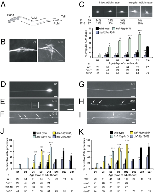
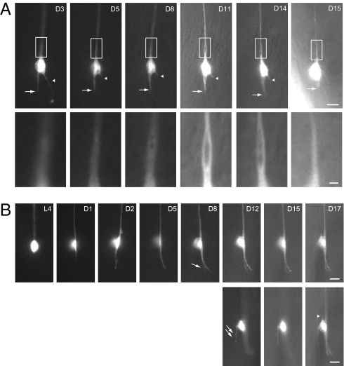
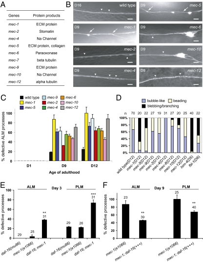
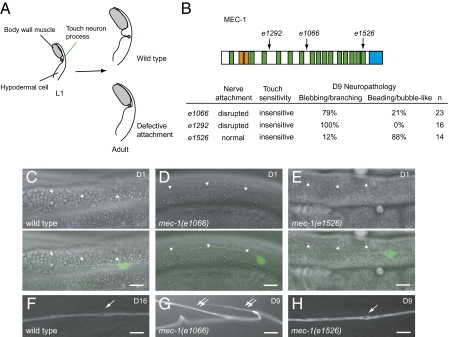
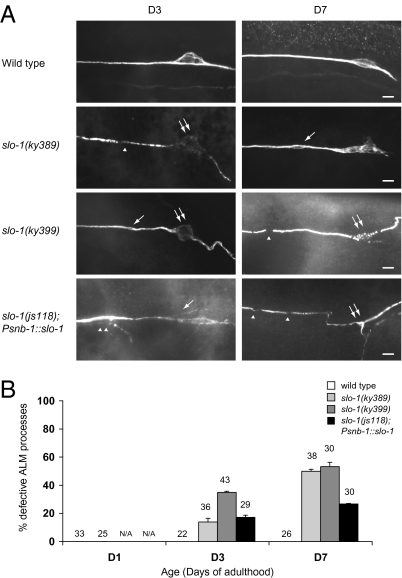
Although many genes have been implicated in the pathogenesis of common neurodegenerative diseases, the genetic and cellular mechanisms that maintain neuronal integrity during normal aging remain elusive. Here we show that Caenorhabditis elegans touch receptor and cholinergic neurons display age-dependent morphological defects, including cytoskeletal disorganization, axon beading, and defasciculation. Progression of neuronal aging is regulated by DAF-2 and DAF-16 signaling, which also modulate adult life span. Mutations that disrupt touch-evoked sensory activity or reduce membrane excitability trigger accelerated neuronal aging, indicating that electrical activity is critical for adult neuronal integrity. Disrupting touch neuron attachment to the epithelial cells induces distinct neurodegenerative phenotypes. These results provide a detailed description of the age-dependent morphological defects that occur in identified neurons of C. elegans, demonstrate that the age of onset of these defects is regulated by specific genes, and offer experimental evidence for the importance of normal levels of neural activity in delaying neuronal aging.


Conflict of interest statement

The authors declare no conflict of interest.

Figures

Fig. 1.
Fig. 1.
Age-dependent defects occur in C. elegans touch receptor neurons. (Scale bar: 5 μm.) (A) Diagram of C. elegans paired ALM and PLM touch receptor neurons (lateral view). Only neurons on the left are shown. (B) Immunostaining of the wild-type ALM neuronal cell bodies with the anti-acetylated α-tubulin antibody 6–11B-1. The age of the animals (days of adulthood) is indicated. (C) Quantification of ALM neurons with irregularly shaped soma in the wild type and the mutants. Representative GFP images of different wild-type ALM neuronal shapes are shown, with number of neurons scored (n) at each time point. GFP is from zdIs5(Pmec-4::gfp). (DI) Epifluorescence images of age-dependent defects of the ALM (D and E) and the PLM (FI) processes in wild type. Anterior is to the left. (E) A bubble-like lesion in the ALM process. Inset is enlarged on the right. Beading (F, arrows), blebbing (H, arrows), and branching (I) of the PLM process are shown. Branching appears to develop from the sites of blebbing (I, arrow). (J and K) Quantification of the ALM (J) and the PLM (K) process abnormalities in wild-type and mutant adult animals. Error bars are SEs of proportions. The number of neurons scored (n) at each time point is provided. *P < 0.05, **P < 0.01, and ***P < 0.001 (Fisher's exact test or two-proportion test).
Fig. 2.
Fig. 2.
Longitudinal imaging of ALM neurons in the wild type. Epifluorescence images of individual ALM neurons at different time points over the animals’ life span (lateral view); anterior is up and ventral side is to the right. GFP is from zdIs5(Pmec-4::gfp). Ages (days of adulthood) are indicated. [Scale bar: 5 μm or 1 μm (A, Insets).] (A) The posterior process of the ALM neuron (arrow), which became slightly shorter and thicker between D3 and D5, remained static from D5 to D14, and retracted on D15. Arrowheads indicate an aberrant protrusion from the cell body, which was truncated between D3 and D5, and completely disappeared on D15. Insets highlight the development of a bubble-like structure at the proximal segment of the ALM process. The animal died on D16. (B) The posterior process of the animal grew out on D1, continued to extend between D1 and D5, and branched at D8 (single arrow). An aberrant protrusion developed from the dorsal side of the cell body at D12 (double arrows). On D17, another short protrusion developed at the anterior pole of the ALM neuron (arrowhead). (Lower) The three images were taken from different focal planes compared with their temporal counterparts in the Upper.
Fig. 3.
Fig. 3.
Progressive touch neuron defects in the mec mutants. (A) Summary of the mec genes and their encoded proteins. (B) Representative live GFP images (except for that of the mec-12 mutant, which is immunofluorescence image-labeled by 6–11B-1) of the ALM processes in the wild type (D16) and the mutants (D9). (Scale bar: 5 μm.) Arrows and arrowheads indicate branching and bubble-like lesions, respectively. (C) Quantification of defective ALM processes. Total numbers of ALM neurons scored (D1/D9/D12): wild type, 29/54/70; mec-1, 25/23/20; mec-2, 17/28/19; mec-4, 24/18/31; mec-5, 31/23/22; mec-6, 19/25/27; mec-9, 20/16/27; mec-10, 23/29/20; mec-12, 19/27/25. (D) Percentages of bubble-like lesions, beading, or blebbing/branching in all of the abnormal events found in the ALM neurons of wild type and the mutants. The age of the animals is specified. (E) Defects of the ALM and the PLM processes of D3 mec-1(e1066) mutants are enhanced by a daf-16(mu86) mutation. (F) Overexpression of DAF-16 from the endogenous daf-16 promoter significantly suppresses ALM and PLM defects of D9 mec-1(e1066) mutants. Error bars are SEs of proportions. **P < 0.01 or ***P < 0.001 (Fisher's exact test or two-proportion test).
Fig. 4.
Fig. 4.
Disruption of nerve attachment or neuronal activity causes distinctive degeneration phenotypes. (Scale bar: 10 μm.) (A) Diagram of touch neuron attachment (transverse section). In wild-type L1 larvae, the process of the touch receptor neuron lies adjacent to the body-wall muscles. The nerve process later adopts the adult position where it is attached to the overlying cuticle and is thus separated from the muscles. In mutants with defective attachment, the touch neuron process remains in the juvenile position and is not attached to the epithelium. (B) MEC-1 protein structure. e1066, splice junction mutation; e1292, nonsense mutation; e1526, missense mutation. Orange, EGF domains; green, Kunitz domains; blue, the C terminus domain. Percentages of the predominant type defects (branching/blebbing or bubble-like/beading) of all axonal defects are provided. (CE) Attachment of the ALM process in wild-type (C) and mec-1 mutants (D and E). (Upper panels) Differential interference contrast (DIC) images. (Lower panels) Overlay of DIC and GFP images. The lower borders of body-wall muscles are indicated by arrowheads. (FH) Representative pathology of aging ALM neurons in wild-type (F) and mec-1 mutants (G and H). Single arrows: bubble-like lesions. Double arrows: branching of the nerve processes.
Fig. 5.
Fig. 5.
Suppression of electrical activity by slo-1 gain-of-function mutations induces progressive touch receptor neuron defects. (Scale bar: 5 μm.) (A) Immunofluorescence images of ALM neurons in the wild-type, slo-1(ky389), slo-1(ky399), and slo-1(js118); Psnb-1::slo-1 animals at D3 and D7 of adulthood, with touch receptor neurons labeled by the 6–11B-1 antibody. ky389 and ky399 are gain-of-function mutations, and js118 is a recessive loss-of-function mutation. Acetylated tubulin was decreased and disorganized in the neuronal cell bodies in slo-1(ky389), slo-1(ky399), and slo-1(js118); Psnb-1::slo-1 mutants. Single arrows: bubble-like lesions; single arrowheads: discontinuity of the acetylated tubulin immunoreactivity; double arrows: ALM cell bodies; double arrowheads: branching from the nerve processes. (B) Quantification of ALM defects in wild type, slo-1(ky389), slo-1(ky399), and slo-1(js118); Psnb-1::slo-1 animals at D1, D3, and D7. Error bars are SEs of proportions. The number of cells scored is indicated. N/A, not available.

References

    1. Yankner BA, Lu T, Loerch P. The aging brain. Annu Rev Pathol. 2008;3:41–66. - PubMed
    1. Garigan D, et al. Genetic analysis of tissue aging in Caenorhabditis elegans: A role for heat-shock factor and bacterial proliferation. Genetics. 2002;161:1101–1112. - PMC - PubMed
    1. Herndon LA, et al. Stochastic and genetic factors influence tissue-specific decline in ageing C. elegans. Nature. 2002;419:808–814. - PubMed
    1. Haithcock E, et al. Age-related changes of nuclear architecture in Caenorhabditis elegans. Proc Natl Acad Sci USA. 2005;102:16690–16695. - PMC - PubMed
    1. Collins JJ, Huang C, Hughes S, Kornfeld K. The measurement and analysis of age-related changes in Caenorhabditis elegans. WormBook. 2008;, Jan 24:1–21. - PMC - PubMed

Publication types