. 2011 May 24;108(21):8565-70.
doi: 10.1073/pnas.1011688108. Epub 2011 May 13.

Imaging the electromechanical activity of the heart in vivo

Affiliations

Imaging the electromechanical activity of the heart in vivo

Jean Provost et al. Proc Natl Acad Sci U S A. .

Abstract

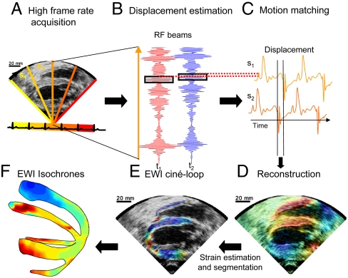
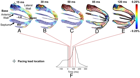
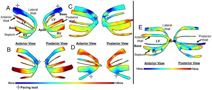
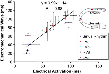
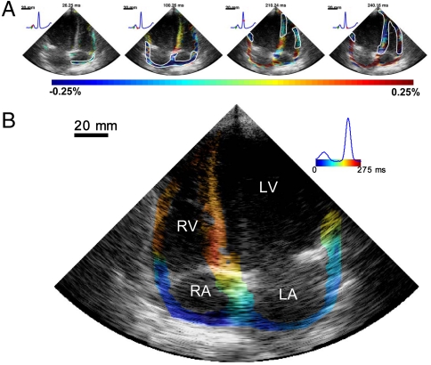
Cardiac conduction abnormalities remain a major cause of death and disability worldwide. However, as of today, there is no standard clinical imaging modality that can noninvasively provide maps of the electrical activation. In this paper, electromechanical wave imaging (EWI), a novel ultrasound-based imaging method, is shown to be capable of mapping the electromechanics of all four cardiac chambers at high temporal and spatial resolutions and a precision previously unobtainable in a full cardiac view in both animals and humans. The transient deformations resulting from the electrical activation of the myocardium were mapped in 2D and combined in 3D biplane ventricular views. EWI maps were acquired during five distinct conduction configurations and were found to be closely correlated to the electrical activation sequences. EWI in humans was shown to be feasible and capable of depicting the normal electromechanical activation sequence of both atria and ventricles. This validation of EWI as a direct, noninvasive, and highly translational approach underlines its potential to serve as a unique imaging tool for the early detection, diagnosis, and treatment monitoring of arrhythmias through ultrasound-based mapping of the transmural electromechanical activation sequence reliably at the point of care, and in real time.


Conflict of interest statement

The authors declare no conflict of interest.

Figures

Fig. 1.
Fig. 1.
Block diagram of the EWI technique. (A) A full view of the two ventricles is first divided in partially overlapping sectors, which are imaged at separate heartbeats. (B) High-precision displacement estimation between two consecutively acquired RF beams (t1, t2) is then performed using very high-frame-rate RF speckle tracking. (C) A region of the heart muscle, common to two neighboring sectors, is then selected. By comparing the temporally varying displacements measured in neighboring sectors (s1, s2) via a cross-correlation technique, the delay between them is estimated. (D) A full-view cine loop of the displacement overlaid onto the B mode can then be reconstructed with all the sectors in the composite image synchronized. (E) The heart walls are then segmented, and incremental strains are computed to depict the EW. (F) By tracking the onset of the EW, isochrones of the sequence of activation are generated.
Fig. 2.
Fig. 2.
Propagation of the EW when paced from the lateral wall, near the base. Activated regions are traced at (A) 15 ms, (B) 30 ms, (C) 50 ms (D) 85 ms, and (E) 120 ms and indicated on the (F) ECG; 0 ms corresponds to the pacing stimulus. (AC) The EW propagates from the basal part of the lateral wall toward the apex. (D) Note that in the apical region, a transition from lengthening to shortening is observed rather than a transition from thinning to thickening. (DE) In the anterior wall, the EW propagates from both the base and apex. The scale shows interframe strains.
Fig. 3.
Fig. 3.
Isochrones showing the activation sequence under different pacing protocols. (A) Pacing from the basal region of the lateral wall. (B) Pacing from the apex. (C) Pacing from the apical region of the lateral wall. (D) Pacing from the apical region of the right-ventricular wall. (E) Isochrones showing the EW activation sequence during sinus rhythm. The activation sequence exhibits early activation at the median level and late activation at the basal and apical levels. Activation of the right-ventricular wall occurred after the activation of the septal and lateral walls.
Fig. 4.
Fig. 4.
Electrical and electromechanical activation times during the four pacing protocols and sinus rhythm in four different heart segments in the posterior and anterior walls, as indicated in the legend. A strong correlation was observed, with a slope of 0.99.
Fig. 5.
Fig. 5.
Normal sinus rhythm in a healthy volunteer (23 y-old female). (A) The EW first occurred in the right atrium and propagated toward the left atrium (blue). This resulted in prestretching of the ventricles (red). The EW then appeared at the midlevel in the septum and close to the apex in the right-ventricular wall, and then propagated toward the apex and the base. (B) Corresponding isochrones. The earliest activation occurred in the right atrium. In the ventricles, it was possible to identify multiple regions of early activation, namely at the midlevel of the septum, near the apex of the right ventricle, and close to the base in the lateral wall. RA, right atrium; LA, left atrium.

References

    1. Bers DM. Cardiac excitation-contraction coupling. Nature. 2002;415:198–205. - PubMed
    1. Ashikaga H, et al. Transmural dispersion of myofiber mechanics: Implications for electrical heterogeneity in vivo. J Am Coll Card. 2007;49:909–916. - PMC - PubMed
    1. Cordeiro JM, Greene L, Heilmann C, Antzelevitch D, Antzelevitch C. Transmural heterogeneity of calcium activity and mechanical function in the canine left ventricle. Am J Physiol Heart Circ Physiol. 2004;286:H1471–H1479. - PubMed
    1. Zhang X, et al. Noninvasive three-dimensional electrocardiographic imaging of ventricular activation sequence. Am J Physiol Heart Circ Physiol. 2005;289:H2724–H2732. - PubMed
    1. Ramanathan C, Ghanem RN, Jia P, Ryu K, Rudy Y. Noninvasive electrocardiographic imaging for cardiac electrophysiology and arrhythmia. Nat Med. 2004;10:422–428. - PMC - PubMed

Publication types

MeSH terms