. 2011 Jul 1;71(13):4412-22.
doi: 10.1158/0008-5472.CAN-10-4111. Epub 2011 May 13.

HDAC4-regulated STAT1 activation mediates platinum resistance in ovarian cancer

Affiliations

HDAC4-regulated STAT1 activation mediates platinum resistance in ovarian cancer

Euan A Stronach et al. Cancer Res. .

Abstract

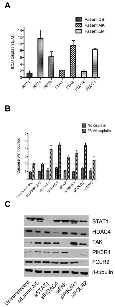
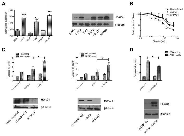
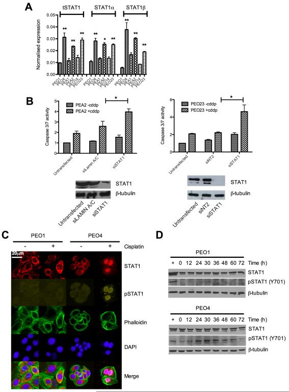
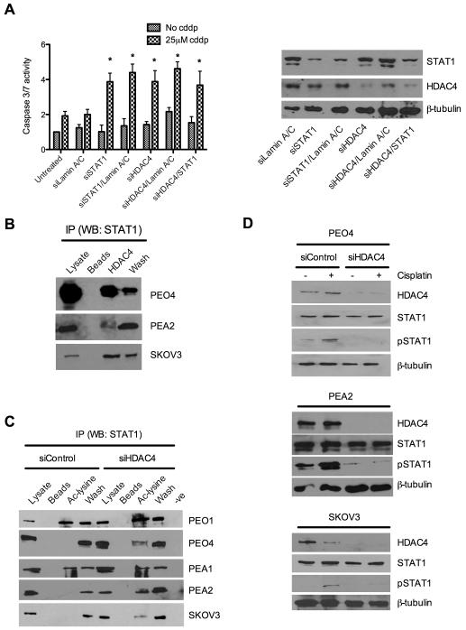
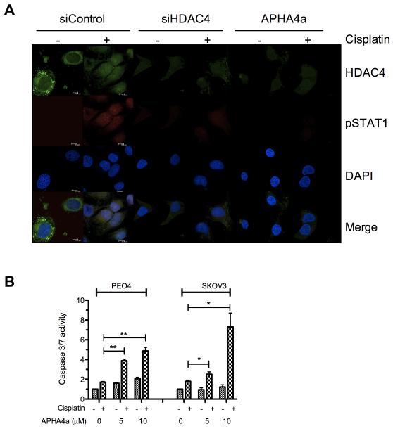
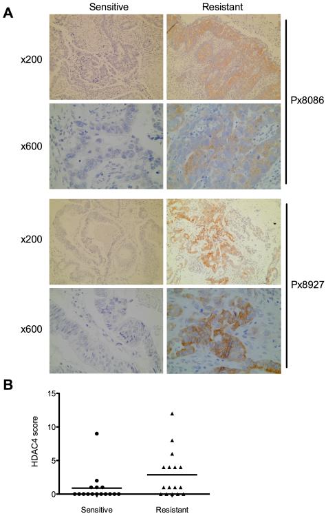
Ovarian cancer frequently acquires resistance to platinum chemotherapy, representing a major challenge for improving patient survival. Recent work suggests that resistant clones exist within a larger drug-sensitive cell population prior to chemotherapy, implying that resistance is selected for rather than generated by treatment. We sought to compare clinically derived, intrapatient paired models of initial platinum response and subsequent resistant relapse to define molecular determinants of evolved resistance. Transcriptional analysis of a matched cell line series from three patients with high-grade serous ovarian cancer before and after development of clinical platinum resistance (PEO1/PEO4/PEO6, PEA1/PEA2, PEO14/PEO23) identified 91 up- and 126 downregulated genes common to acquired resistance. Significantly enhanced apoptotic response to platinum treatment in resistant cells was observed following knockdown of histone deacetylase (HDAC) 4, FOLR2, PIK3R1, or STAT1 (P < 0.05). Interestingly, HDAC4 and STAT1 were found to physically interact. Acetyl-STAT1 was detected in platinum-sensitive cells but not in HDAC4 overexpressing platinum-resistant cells from the same patient. In resistant cells, STAT1 phosphorylation/nuclear translocation was seen following platinum exposure, whereas silencing of HDAC4 increased acetyl-STAT1 levels, prevented platinum-induced STAT1 activation, and restored cisplatin sensitivity. Conversely, matched sensitive cells were refractory to STAT1 phosphorylation on platinum treatment. Analysis of 16 paired tumor biopsies taken before and after development of clinical platinum resistance showed significantly increased HDAC4 expression in resistant tumors [n = 7 of 16 (44%); P = 0.04]. Therefore, clinical selection of HDAC4-overexpressing tumor cells upon exposure to chemotherapy promotes STAT1 deacetylation and cancer cell survival. Together, our findings identify HDAC4 as a novel, therapeutically tractable target to counter platinum resistance in ovarian cancer.


Figures

Figure 1
Figure 1. Identification of platinum resistance modulators
IC50s for platinum sensitive and resistant paired cell lines were calculated based on SRB assay data (n=3). This analysis confirmed an in vitro resistance to cisplatin in all clinically platinum resistant cell lines relative to their sensitive parent lines (Fold changes in IC50 between sensitive and matched resistant cells: PEO4- 8.7 [p=0.016], PEO6- 4.6 [p=0.004], PEA2- 4.1 [p=0.0002], PEO23- 4.5 [p=0.00002]) (A). Following microarray based expression analysis candidate genes were assessed for a role in acquired platinum resistance by a first round of siRNA knockdown followed by cisplatin treatment identifying six genes that enhanced apoptotic induction by cisplatin. Following replication, four of these genes (FOLR2 [p=0.004], HDAC4 [p=0.02], PIK3R1 [p=0.04] and STAT1 [p=0.0002]) passed statistical analysis (see also table 1) as enhancing apoptotic induction on cisplatin treatment when compared to control siRNA treated cells. Data are represented as means ±SEM of 4 replicate experiments (B). Specificity of siRNA was confirmed by western blot (C). All t-tests throughout are two-tailed and assume unequal variances.
Figure 2
Figure 2. HDAC4 contributes to cisplatin resistance
Over-expression of HDAC4 in platinum resistant cells was confirmed at the RNA (left) and protein level (right) (A) [***p<0.005 t-test]. The effect of HDAC4 knockdown on cisplatin response was validated by SRB assay, 72hr post cisplatin exposure of siRNA treated PEO4 cells. Data are mean ± SEM (n=3) (B). Clinically derived platinum resistant cell lines (PEA2/PEO23) were assessed for the contribution of HDAC4 to acquired resistance. Data in the upper panes represent the mean caspase 3/7 induction relative to control siRNA ±SEM (n=3) whereas lower pane shows confirmation of HDAC4 knockdown by western blot. *p<0.05 t-test comparing caspase 3/7 activation between siRNA treated and siRNA control cells following cisplatin exposure (C). Over-expression of HDAC4 in the platinum sensitive PEO1 cell line as described in the supplementary methods. Over-expression of protein (lower) was confirmed by western blot and accompanying caspase activation data (upper) demonstrates attenuated apoptotic response to cisplatin in cells over-expressing HDAC4 (n=4; p=0.032) comparing caspase 3/7 activation between pcDNA3.1-empty vector (EV) transfected and pcDNA3.1-HDAC4 treated cells (D).
Figure 3
Figure 3. STAT1 contributes to cisplatin resistance and is phosphorylated at Y701 in resistant cells following platinum treatment
cDNA from sensitive and resistant cell lines was quantified for expression of total STAT1 and the α and β isoforms of STAT1 individually (A) [*p<0.05, **p<0.01 t-tests comparing resistant cells to their sensitive matched line]. siRNA to STAT1 in further platinum resistant cell lines revealed a general effect of resensitisation to cisplatin following STAT1 knockdown as measured by caspase 3/7 induction. Effective knockdown was confirmed by western blot (See also supplementary figure S5) *p<0.05 t-test comparing caspase 3/7 activation between siRNA treated and siRNA control cells following cisplatin exposure (B). Subcellular location of total- and phospho-STAT1 was determined in sensitive and resistant cells by immunofluorescence microscopy as described. Nuclei were revealed using DAPI stain and actin cytoskeleton visualised by Alexa 633nm conjugated phalloidin stain (C) (see also supplementary figure S6). Identical matched cells were treated with cisplatin over 72hrs at 5μM for sensitive PEO1 cells and 25μM for resistant PEO4. Protein lysates were collected and total and phospho-STAT1 determined by western blot (D).
Figure 4
Figure 4. HDAC4 and STAT1 act in a single, novel pathway in which HDAC4 is required for deacetylation and phosphorylation of STAT1 in response to cisplatin
STAT1 and HDAC4 were knocked down alone and in combination using siRNA and induction of caspase 3/7 assessed ±cisplatin treatment in platinum resistant PEO4 cells *p<0.05 t-test comparing caspase 3/7 activation between siRNA treated and siRNA control cells following cisplatin exposure (left). Western blot confirmed knockdown at protein level (right) (A). Immunoprecipitates from untreated platinum resistant PEO4, PEA2 and SKOV3 cells were made using anti-HDCA4 antibody and probed for the presence of STAT1 (B). Immunoprecipitates from the platinum sensitive cell lines PEO1 and PEA1, the resistant paired lines PEO4 and PEA2 and the resistant line SKOV-3 were prepared using anti-acetyl lysine antibody and probed for the presence of STAT1 both before and after siRNA based knockdown of HDAC4 (C). Control and HDAC4 siRNA treated PEO4 (top), PEA2 (middle) and SKOV3 (bottom) cell lysates ± cisplatin were probed for phosphorylation of STAT1 at tyrosine 701. (D). Legend for (B) and (C): lysates- whole cell extract pre-immunoprecipitation; beads- protein G sepharose beads without primary antibody; HDAC4/Ac-lysine- specific immunoprecipitates; wash-whole-cell lysate column flow-through; −ve - primary antibody/lysate without protein G sepharose beads.
Figure 5
Figure 5. HDAC inhibition prevents nuclear localisation of phospho-STAT1 and resensitises resistant cells to cisplatin
Immuno-fluorescent microscopy of platinum resistant SKOV3 cells reveals induction and nuclear localisation of pSTAT1-Y701 on 24hrs stimulation with 25μM cisplatin and shows that HDAC4 knockdown using siRNA or inhibition using 5μM APHA4a prevents accumulation of pSTAT1 (A). Treatment of the resistant cells PEO4 or SKOV3 with the HDAC inhibitor APHA4a at 5μM or 10μM enhances the apoptotic response to treatment with 25μM cisplatin measured after 24hr incubation (B) *p<0.05, **p<0.01 t-test comparing caspase 3/7 activation between vehicle and APHA4a treated cells following cisplatin exposure.
Figure 6
Figure 6. HDAC4 expression increases on acquisition of cisplatin resistance in matched clinical specimens
Sections cut from FFPE ovarian tumor blocks from before and after acquisition of platinum resistance in individual patients were stained for HDAC4 expression and intensity and frequency of staining. Photomicrographs indicate increased expression of HDAC4 (brown staining) in resistant (right) compared to sensitive (left) biopsies from the same patient. Magnifications are indicated. (A). HDCA4 expression scores were produced for each section as described and the change in score on acquisition of platinum resistance analysed by Wilcoxon rank sum test. Scores for 16 paired sections, grouped as sensitive and resistant are indicated (p=0.0413) (B).

References

    1. Herzog TJ. Recurrent ovarian cancer: how important is it to treat to disease progression? Clin Cancer Res. 2004;10:7439–49. - PubMed
    1. Agarwal R, Kaye SB. Ovarian cancer: strategies for overcoming resistance to chemotherapy. Nat Rev Cancer. 2003;3:502–16. - PubMed
    1. Kartalou M, Essigmann JM. Mechanisms of resistance to cisplatin. Mutat Res. 2001;478:23–43. - PubMed
    1. Rabik CA, Dolan ME. Molecular mechanisms of resistance and toxicity associated with platinating agents. Cancer Treat Rev. 2007;33:9–23. - PMC - PubMed
    1. Cooke SL, Ng CKY, Melnyk N, Garcia MJ, Hardcastle T, Temple J, et al. Genomic analysis of genetic heterogeneity and evolution in high-grade serous ovarian carcinoma. Oncogene. 2010;29:4905–13. - PMC - PubMed

Publication types

MeSH terms