

Flow cytometric immunophenotypic assessment of T-cell clonality by Vβ repertoire analysis: detection of T-cell clonality at diagnosis and monitoring of minimal residual disease following therapy
- PMID: 21571962
- PMCID: PMC7384354
- DOI: 10.1309/AJCPV2D1DDSGJDBW
Flow cytometric immunophenotypic assessment of T-cell clonality by Vβ repertoire analysis: detection of T-cell clonality at diagnosis and monitoring of minimal residual disease following therapy
Abstract
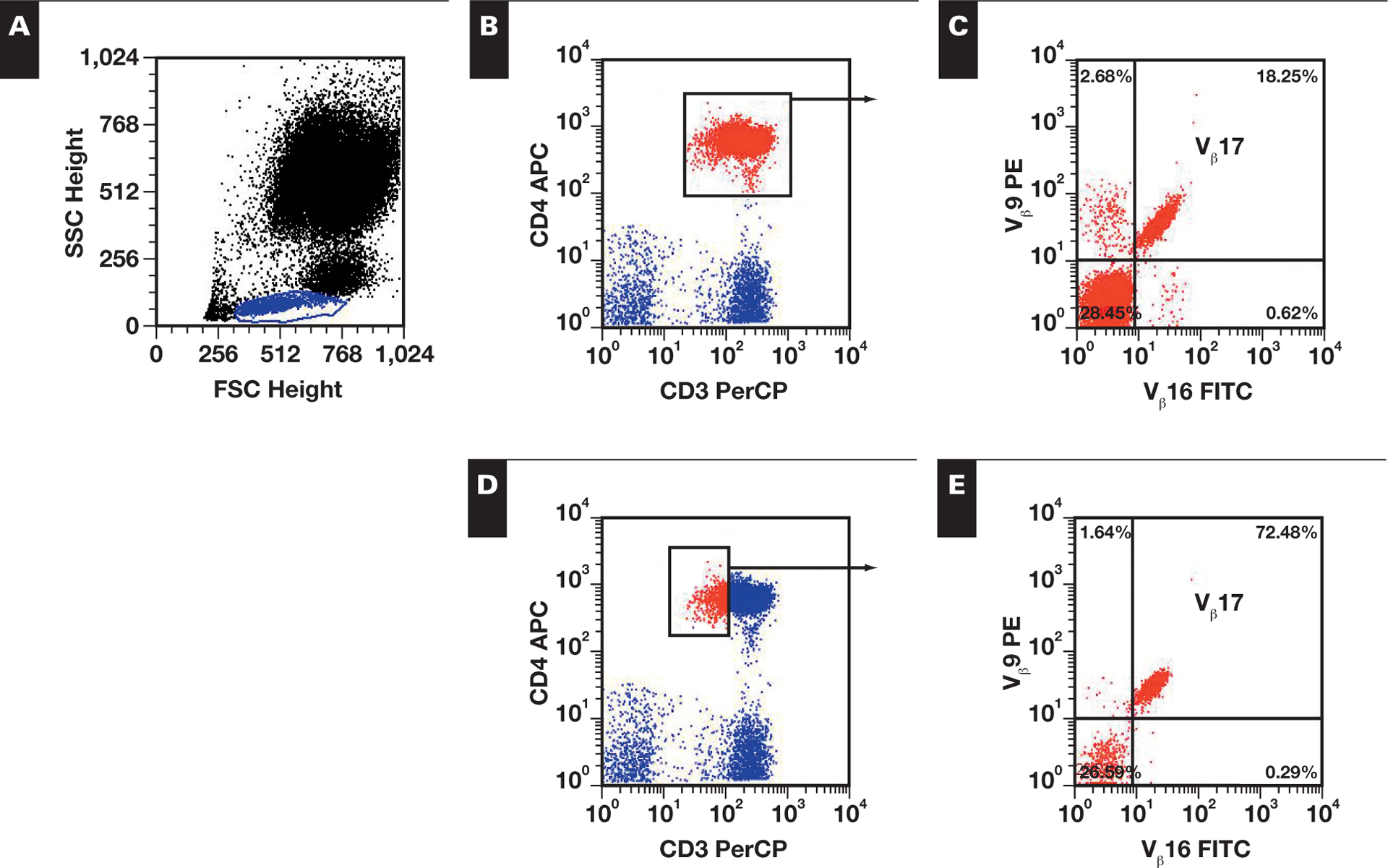
Flow cytometric T-cell receptor (TCR)-V(β) repertoire analysis (TCR-V(β)-R) is a sensitive method for detection of T-cell clonality; however, no uniform approach exists to define clonality in neoplastic T cells. TCR-V(β)-R was evaluated in patients with a diagnosis of T-cell neoplasia in initial diagnostic specimens from 41 patients and for minimal residual disease (MRD) monitoring in 61 sequential samples from 14 patients with mature T-cell neoplasia. Gating strategies and criteria for detection of T-cell clonality were determined. In all 41 initial specimens, T-cell clonality was demonstrated via TCR-V(β)-R. The frequency of V(β) usage was consistent with random neoplastic transformation of TCR-V(β) subsets. MRD was successfully detected in follow-up samples from all 14 patients evaluated, Furthermore, MRD after therapy was quantitated in 48 peripheral blood specimens. TCR-V(β)-R analysis is a sensitive method for detection of T-cell clonality and is useful for diagnosis and MRD detection in multiple specimen types.
Figures
References
-
- Rawstron AC, Villamor N, Ritgen M, et al. International standardized approach for flow cytometric residual disease monitoring in chronic lymphocytic leukaemia. Leukemia. 2007;21:956–964. - PubMed
-
- Rawstron AC, Kennedy B, Evans PAS, et al. Quantitation of minimal disease levels in chronic lymphocytic leukemia using a sensitive flow cytometric assay improves the prediction of outcome and can be used to optimize therapy. Blood. 2001;98:29–35. - PubMed
-
- Sausville J, Salloum RG, Sorbara L, et al. Minimal residual disease detection in hairy cell leukemia: comparison of flow cytometric immunophenotyping with clonal analysis using consensus primer polymerase chain reaction for the heavy chain gene. Am J Clin Pathol. 2003;119:213–217. - PubMed
-
- Stetler-Stevenson MA, Ahmad E, Barnett D, et al. Clinical Flow Cytometric Analysis of Neoplastic Hematolymphoid Cells; Approved Guideline 2nd ed Wayne, PA: Clinical and Laboratory Standards Institute; 2005. CLSI document H43–A2.
Publication types
MeSH terms
Substances
Grants and funding
LinkOut - more resources
Full Text Sources
Other Literature Sources
