

Doppler velocity measurements from large and small arteries of mice
- PMID: 21572013
- PMCID: PMC3154663
- DOI: 10.1152/ajpheart.00320.2011
Doppler velocity measurements from large and small arteries of mice
Abstract
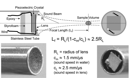
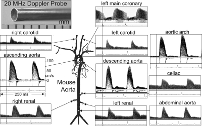
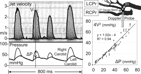
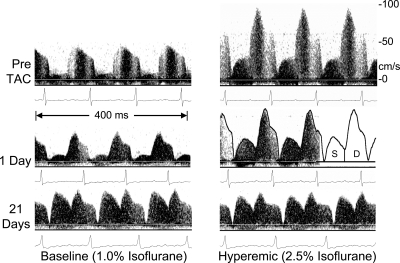
With the growth of genetic engineering, mice have become increasingly common as models of human diseases, and this has stimulated the development of techniques to assess the murine cardiovascular system. Our group has developed nonimaging and dedicated Doppler techniques for measuring blood velocity in the large and small peripheral arteries of anesthetized mice. We translated technology originally designed for human vessels for use in smaller mouse vessels at higher heart rates by using higher ultrasonic frequencies, smaller transducers, and higher-speed signal processing. With these methods one can measure cardiac filling and ejection velocities, velocity pulse arrival times for determining pulse wave velocity, peripheral blood velocity and vessel wall motion waveforms, jet velocities for the calculation of the pressure drop across stenoses, and left main coronary velocity for the estimation of coronary flow reserve. These noninvasive methods are convenient and easy to apply, but care must be taken in interpreting measurements due to Doppler sample volume size and angle of incidence. Doppler methods have been used to characterize and evaluate numerous cardiovascular phenotypes in mice and have been particularly useful in evaluating the cardiac and vascular remodeling that occur following transverse aortic constriction. Although duplex ultrasonic echo-Doppler instruments are being applied to mice, dedicated Doppler systems are more suitable for some applications. The magnitudes and waveforms of blood velocities from both cardiac and peripheral sites are similar in mice and humans, such that much of what is learned using Doppler technology in mice may be translated back to humans.
Figures

References
-
- Aristizabal O, Christopher DA, Foster FS, Turnbull DH. 40-MHz echocardiographic scanner for cardiovascular assessment of mouse embryos. Ultrasound Med Biol 24: 1407–1417, 1998 - PubMed
-
- Asmar R, Benetos A, Topouchian J, Laurent P, Pannier B, Brisac AM, Target R, Levy BI. Assessment of arterial distensibility by automatic pulse wave velocity measurement. Validation and clinical studies. Hypertension 26: 485–490, 1995 - PubMed
-
- Baker DW. Pulsed ultrasonic Doppler blood flow sensing. IEEE Trans Sonics Ultrason SU-17: 170–185, 1970
-
- Bonnin P, Sabaa N, Flamant M, Debbabi H, Tharaux PL. Ultrasound imaging of renal vaso-occlusive events in transgenic sickle mice exposed to hypoxic stress. Ultrasound Med Biol 34: 1076–1084, 2008 - PubMed
-
- Bonnin P, Villemain A, Vincent F, Debbabi H, Silvestre JS, Contreses JO, Levy BI, Tobelem G, Dupuy E. Ultrasonic assessement of hepatic blood flow as a marker of mouse hepatocarcinoma. Ultrasound Med Biol 33: 561–570, 2007 - PubMed
Publication types
MeSH terms
Grants and funding
- HL-89792/HL/NHLBI NIH HHS/United States
- HL-57068/HL/NHLBI NIH HHS/United States
- HL-42267/HL/NHLBI NIH HHS/United States
- AG-15568/AG/NIA NIH HHS/United States
- R01 HL022512/HL/NHLBI NIH HHS/United States
- HL-42313/HL/NHLBI NIH HHS/United States
- P41 RR011795/RR/NCRR NIH HHS/United States
- HL-52364/HL/NHLBI NIH HHS/United States
- P41 EB002182/EB/NIBIB NIH HHS/United States
- R01 HL089792/HL/NHLBI NIH HHS/United States
- HL-13870/HL/NHLBI NIH HHS/United States
- HL-42550/HL/NHLBI NIH HHS/United States
- AG-13251/AG/NIA NIH HHS/United States
- HL-22512/HL/NHLBI NIH HHS/United States
LinkOut - more resources
Full Text Sources
Other Literature Sources