

Electromechanical models of the ventricles
- PMID: 21572017
- PMCID: PMC3154669
- DOI: 10.1152/ajpheart.00324.2011
Electromechanical models of the ventricles
Abstract
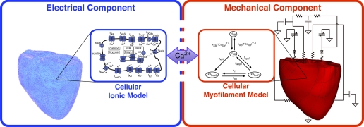
Computational modeling has traditionally played an important role in dissecting the mechanisms for cardiac dysfunction. Ventricular electromechanical models, likely the most sophisticated virtual organs to date, integrate detailed information across the spatial scales of cardiac electrophysiology and mechanics and are capable of capturing the emergent behavior and the interaction between electrical activation and mechanical contraction of the heart. The goal of this review is to provide an overview of the latest advancements in multiscale electromechanical modeling of the ventricles. We first detail the general framework of multiscale ventricular electromechanical modeling and describe the state of the art in computational techniques and experimental validation approaches. The powerful utility of ventricular electromechanical models in providing a better understanding of cardiac function is then demonstrated by reviewing the latest insights obtained by these models, focusing primarily on the mechanisms by which mechanoelectric coupling contributes to ventricular arrythmogenesis, the relationship between electrical activation and mechanical contraction in the normal heart, and the mechanisms of mechanical dyssynchrony and resynchronization in the failing heart. Computational modeling of cardiac electromechanics will continue to complement basic science research and clinical cardiology and holds promise to become an important clinical tool aiding the diagnosis and treatment of cardiac disease.
Figures
Similar articles
-
Spatial velocity of the dynamic vectorcardiographic loop provides crucial insight in ventricular dysfunction.Med Hypotheses. 2020 Feb;135:109484. doi: 10.1016/j.mehy.2019.109484. Epub 2019 Nov 11. Med Hypotheses. 2020. PMID: 31739078
-
Biomechanics of cardiac electromechanical coupling and mechanoelectric feedback.J Biomech Eng. 2014 Feb;136(2):021007. doi: 10.1115/1.4026221. J Biomech Eng. 2014. PMID: 24337452 Free PMC article. Review.
-
Modeling cardiac electromechanics and mechanoelectrical coupling in dyssynchronous and failing hearts: insight from adaptive computer models.J Cardiovasc Transl Res. 2012 Apr;5(2):159-69. doi: 10.1007/s12265-012-9346-y. Epub 2012 Jan 21. J Cardiovasc Transl Res. 2012. PMID: 22271009 Free PMC article. Review.
-
Development, calibration, and validation of a novel human ventricular myocyte model in health, disease, and drug block.Elife. 2019 Dec 24;8:e48890. doi: 10.7554/eLife.48890. Elife. 2019. PMID: 31868580 Free PMC article.
-
Models of cardiac electromechanics based on individual hearts imaging data: image-based electromechanical models of the heart.Biomech Model Mechanobiol. 2011 Jun;10(3):295-306. doi: 10.1007/s10237-010-0235-5. Epub 2010 Jun 30. Biomech Model Mechanobiol. 2011. PMID: 20589408 Free PMC article.
Cited by
-
Numerical quadrature and operator splitting in finite element methods for cardiac electrophysiology.Int J Numer Method Biomed Eng. 2013 Nov;29(11):1243-66. doi: 10.1002/cnm.2573. Epub 2013 Jul 19. Int J Numer Method Biomed Eng. 2013. PMID: 23873868 Free PMC article.
-
High Spatial Resolution Multi-Organ Finite Element Modeling of Ventricular-Arterial Coupling.Front Physiol. 2018 Mar 2;9:119. doi: 10.3389/fphys.2018.00119. eCollection 2018. Front Physiol. 2018. PMID: 29551977 Free PMC article.
-
Mathematical modeling and simulation of ventricular activation sequences: implications for cardiac resynchronization therapy.J Cardiovasc Transl Res. 2012 Apr;5(2):146-58. doi: 10.1007/s12265-011-9343-6. Epub 2012 Jan 27. J Cardiovasc Transl Res. 2012. PMID: 22282106 Free PMC article. Review.
-
The Living Heart Project: A robust and integrative simulator for human heart function.Eur J Mech A Solids. 2014 Nov;48:38-47. doi: 10.1016/j.euromechsol.2014.04.001. Eur J Mech A Solids. 2014. PMID: 25267880 Free PMC article.
-
Organ-level validation of a cross-bridge cycling descriptor in a left ventricular finite element model: effects of ventricular loading on myocardial strains.Physiol Rep. 2017 Nov;5(21):e13392. doi: 10.14814/phy2.13392. Physiol Rep. 2017. PMID: 29122952 Free PMC article.
References
-
- Guccione J, Costa K, McCulloch A. Finite element stress analysis of left ventricular mechanics in the beating dog heart. J Biomech 28: 1167–1177, 1995 - PubMed
Publication types
MeSH terms
Grants and funding
LinkOut - more resources
Full Text Sources
Medical