. 2011 Aug;301(2):H331-43.
doi: 10.1152/ajpheart.00131.2011. Epub 2011 May 13.

Mechanical control of cation channels in the myogenic response

Affiliations

Mechanical control of cation channels in the myogenic response

Brian E Carlson et al. Am J Physiol Heart Circ Physiol. 2011 Aug.

Abstract

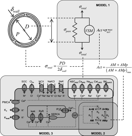
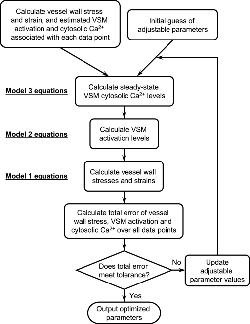
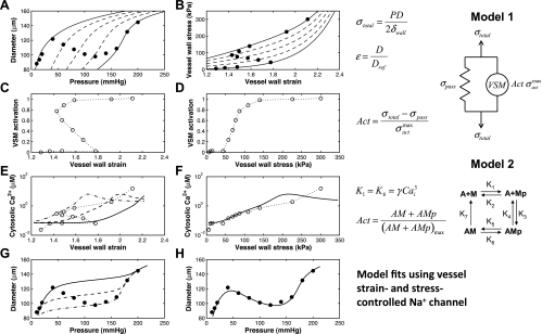
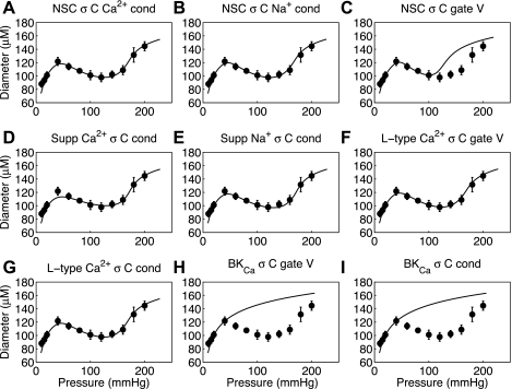
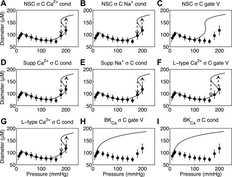
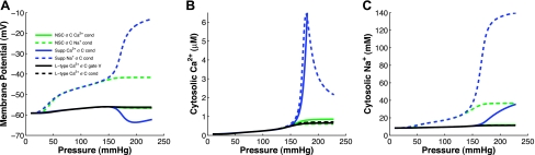
Microcirculatory vessel response to changes in pressure, known as the myogenic response, is a key component of a tissue's ability to regulate blood flow. Experimental studies have not clearly elucidated the mechanical signal in the vessel wall governing steady-state reduction in vessel diameter upon an increase in intraluminal pressure. In this study, a multiscale computational model is constructed from established models of vessel wall mechanics, vascular smooth muscle (VSM) force generation, and VSM Ca(2+) handling and electrophysiology to compare the plausibility of vessel wall stress or strain as an effective mechanical signal controlling steady-state vascular contraction in the myogenic response. It is shown that, at the scale of a resistance vessel, wall stress, and not stretch (strain), is the likely physiological signal controlling the steady-state myogenic response. The model is then used to test nine candidate VSM stress-controlled channel variants by fitting two separate sets of steady-state myogenic response data. The channel variants include nonselective cation (NSC), supplementary Ca(2+) and Na(+), L-type Ca(2+), and large conductance Ca(2+)-activated K(+) channels. The nine variants are tested in turn, and model fits suggest that stress control of Ca(2+) or Na(+) influx through NSC, supplementary Ca(2+) or Na(+), or L-type Ca(2+) channels is sufficient to produce observed steady-state diameter changes with pressure. However, simulations of steady-state VSM membrane potential, cytosolic Ca(2+), and Na(+) with pressure show only that Na(+) influx through NSC channel also generates known trends with increasing pressure, indicating that stress-controlled Na(+) influx through NSC is sufficient to generate the myogenic response.


Figures

Fig. 1.
Fig. 1.
Integrated multiscale model of myogenic response in microcirculatory vessels showing 3 submodels. Model 1: steady state vessel wall mechanics model (4). Model 2: 4-state vascular smooth muscle (VSM) cross-bridge model (14, 24). Model 3: VSM electrophysiology model (22) with vessel stress-controlled channels inserted (crosshatched). Vessel wall stress determines steady-state cytosolic Ca2+ levels in VSM that governs VSM activation and in turn defines the vessel diameter. See methods for details of each of these integrated models.
Fig. 2.
Fig. 2.
Optimization protocol for adjustable parameters to produce best fit of model to experimental data. Vessel wall stress, strain, and estimates of VSM activation and cytosolic Ca2+ are calculated for each data point. Initial guesses of the adjustable parameters are made to simulate the cytosolic Ca2+ concentration as a function of the mechanical stimulus using model 3. Model 2 and model 1 equations are then used to simulate VSM activations and vessel wall stresses and strains. The total error between calculated and simulated values is determined and if error tolerance is not met then the adjustable parameter values are updated and the simulation process is repeated.
Fig. 3.
Fig. 3.
Comparison between vessel strain- and vessel stress-controlled channel functions in the myogenic response. A and B: experimental data of a Wistar-Kyoto (control) rat mesenteric myogenic response from Fig. 1 of Bund (3) given in diameter-pressure (A) and stress-strain (B) domains. Strain is calculated with respect to an estimated reference passive vessel diameter at 5 mmHg. Model 1 is used to draw lines of constant VSM activation (0, 0.25, 0.5, 0.75, and 1) to show levels of activation at each data point. The 2 panels of the second row show the activation level calculated from model 1 as a function of vessel strain (C) or vessel stress (D). The third row of panels uses model 2 to estimate the cytosolic Ca2+ concentration at each data point, which is again plotted against vessel strain (E) and vessel stress (F). Model 2 may overestimate the cytosolic Ca2+ concentration at full VSM activation (last three data points here). The fully integrated model is used to show 3 different fits to Ca2+ as a function of vessel strain with a strain-controlled supplementary Na+ channel inserted into the model (E) and a single fit as a function of vessel stress with a stress-controlled supplementary Na+ channel (F). The bottom 2 panels show the corresponding fully integrated model fits in terms of diameter-pressure with strain-controlled (G) and stress-controlled (H) supplementary Na+ channels. Solid data points in this figure refer to measured experimental data whereas open data points are estimated values calculated by using model 1 and model 2 portions of the fully integrated model.
Fig. 4.
Fig. 4.
Fits to Wistar-Kyoto (control) rat mesenteric arteriole myogenic response from Bund (3) for 9 different proposed vessel stress-controlled channel variants. The 9 variants are nonselective cation (NSC) with stress-controlled (σC) Ca2+ conductance (cond) (A), NSC with stress-controlled Na+ conductance (B), NSC with stress-controlled gating voltage (gate V) (C), supplementary (Supp) Ca2+ with stress-controlled conductance (D), supplementary Na+ with stress-controlled conductance (E), L-type Ca2+ with stress-controlled gating voltage (F), L-type Ca2+ with stress-controlled conductance (G), large-conductance, Ca2+-activated K+ (BKCa) with stress-controlled gating voltage (H), and BKCa with stress-controlled conductance (I).
Fig. 5.
Fig. 5.
Fits to Wistar-Kyoto (control) rat femoral arteriole myogenic response from Bund (3) for 9 different proposed vessel stress-controlled channel variants. Panels are arranged as in Fig. 4. Hysteresis in steady-state response predicted at high pressure for increasing and decreasing pressure steps has been previously documented experimentally and described physiologically (38).
Fig. 6.
Fig. 6.
Simulated steady-state membrane potential (A), cytosolic Ca2+ (B), and Na+ (C) with increasing intraluminal pressure for 6 of the vessel stress-controlled channel formulations that successfully fit the myogenic response data. Only the stress-controlled conductance of the NSC Na+ and supplementary Na+ show significant depolarization over the pressure range simulated. However, the cytosolic Ca2+ concentration with a stress-controlled supplementary Na+ conductance reaches levels in the μM range and cytosolic Na+ essentially equilibrates with extracellular Na+ concentrations both of which are outside of physiological levels.

Similar articles

Cited by

References

    1. Baek EB, Jin C, Park SJ, Park KS, Yoo HY, Jeon JH, Earm YE, Kim SJ. Differential recruitment of mechanisms for myogenic responses according to luminal pressure and arterial types. Pflügers Arch 460: 19–29, 2010 - PubMed
    1. Bayliss WM. On the local reactions of the arterial wall to changes of internal pressure. J Physiol 28: 220–231, 1902 - PMC - PubMed
    1. Bund SJ. Spontaneously hypertensive rat resistance artery structure related to myogenic and mechanical properties. Clin Sci (Lond) 101: 385–393, 2001 - PubMed
    1. Carlson BE, Secomb TW. A theoretical model for the myogenic response based on the length-tension characteristics of vascular smooth muscle. Microcirculation 12: 327–338, 2005 - PubMed
    1. Chen J, Sgouralis I, Moore LC, Layton HE, Layton AT. A mathematical model of the myogenic response to systolic pressure in the afferent arteriole. Am J Physiol Renal Physiol 300: F669–F681, 2011 - PMC - PubMed

Publication types

LinkOut - more resources