

Focal central white matter lesions in Alexander disease
- PMID: 21572052
- PMCID: PMC4154515
- DOI: 10.1177/0883073811405381
Focal central white matter lesions in Alexander disease
Abstract
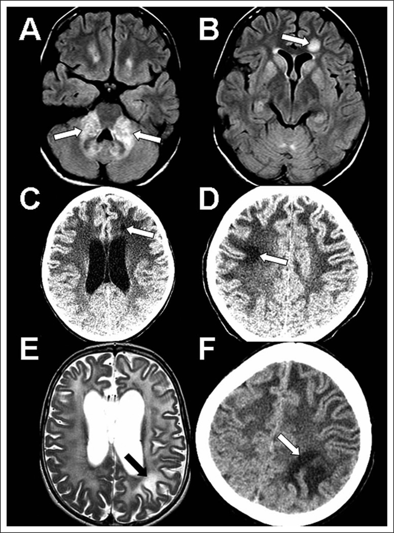
Alexander disease is a neurodegenerative disorder of the central white matter caused by dominant mutations in GFAP, the gene encoding glial fibrillary acidic protein. Magnetic resonance imaging pattern recognition studies have established characteristic radiologic phenotypes for this disorder. In some cases, however, genetically confirmed cases do not express these features, and several reports have identified "atypical" radiologic findings in Alexander disease patients. Here, the authors report 3 genetically confirmed Alexander disease cases with focal central white matter lesions that, upon longitudinal clinical and radiologic evaluation, appear to reflect an atypical Alexander disease magnetic resonance imaging phenotype and not another pathophysiologic process such as encephalitis, infarction, or neoplasm.
Conflict of interest statement
The authors declared no potential conflicts of interest with respect to the research, authorship, and/or publication of this article.
Figures
References
-
- Brenner M, Johnson AB, Boespflug-Tanguy O, et al. Mutations in GFAP, encoding glial fibrillary acidic protein, are associated with Alexander disease. Nat Genet. 2001;27:117–120. - PubMed
-
- van der Knaap MS, Salomons GS, Li R, et al. Unusual variants of Alexander’s disease. Ann Neurol. 2005;57:327–338. - PubMed
Publication types
MeSH terms
Grants and funding
LinkOut - more resources
Full Text Sources
Miscellaneous
