. 2011 Jun;43(6):590-4.
doi: 10.1038/ng.836. Epub 2011 May 15.

Recessive LAMC3 mutations cause malformations of occipital cortical development

Affiliations

Recessive LAMC3 mutations cause malformations of occipital cortical development

Tanyeri Barak et al. Nat Genet. 2011 Jun.

Abstract

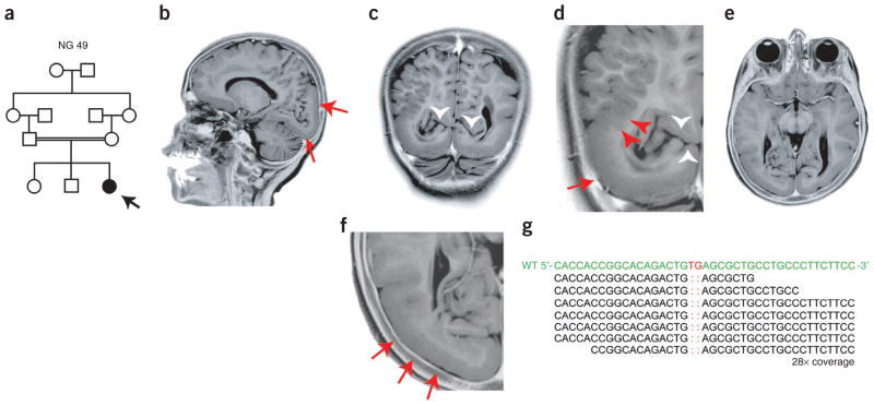
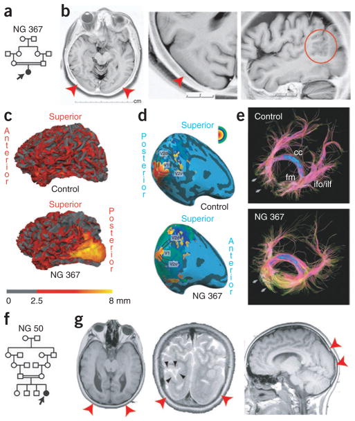
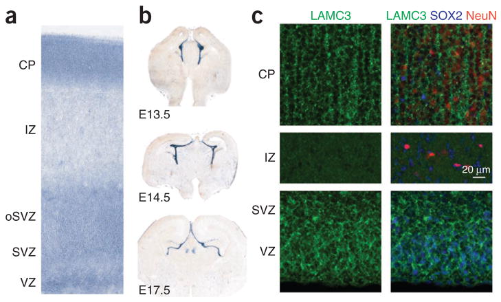
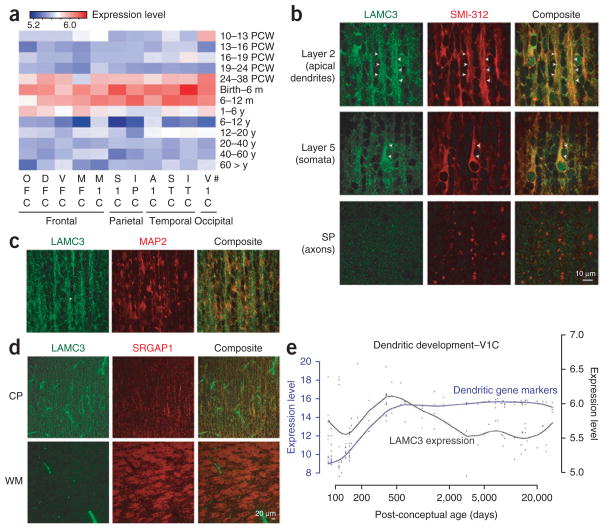
The biological basis for regional and inter-species differences in cerebral cortical morphology is poorly understood. We focused on consanguineous Turkish families with a single affected member with complex bilateral occipital cortical gyration abnormalities. By using whole-exome sequencing, we initially identified a homozygous 2-bp deletion in LAMC3, the laminin γ3 gene, leading to an immediate premature termination codon. In two other affected individuals with nearly identical phenotypes, we identified a homozygous nonsense mutation and a compound heterozygous mutation. In human but not mouse fetal brain, LAMC3 is enriched in postmitotic cortical plate neurons, localizing primarily to the somatodendritic compartment. LAMC3 expression peaks between late gestation and late infancy, paralleling the expression of molecules that are important in dendritogenesis and synapse formation. The discovery of the molecular basis of this unusual occipital malformation furthers our understanding of the complex biology underlying the formation of cortical gyrations.


Conflict of interest statement

COMPETING FINANCIAL INTERESTS

The authors declare competing financial interests: details accompany the full-text HTML version of the paper at http://www.nature.com/naturegenetics/.

Figures

Figure 1
Figure 1
LAMC3 mutation in NG 49 with occipital cortical malformation. (a) The pedigree structure of NG 49 (index case, NG 49-1, black arrow) is shown, which reveals a first cousin marriage. (b) A sagittal magnetic resonance image reveals occipital cortical pachygyria (red arrows). (cf) Coronal (c,d) and axial (e,f) magnetic resonance images reveal thickening and smoothening of the occipital cortex (red arrows) along with polymicrogyria (red arrowheads). Loss of the clear distinction between the gray and white matter is visualized in c and d, in which the white arrowheads mark the preserved calcarine fissure. Panels d and f are enlarged views of c and e, respectively. Photographically inverted T2 images are shown. (g) Exome sequencing shows a 2-bp ‘TG’ deletion (marked in red in the wild-type (WT) sequence on top) in the LAMC3. The depth of coverage across the variant was 28×, and all but one of the reads showed the deletion.
Figure 2
Figure 2
Additional LAMC3 mutations in individuals with occipital cortical malformation. (a) Simplified pedigree structure of NG 367 (arrow, index case) shows a first cousin consanguineous union. (b) Axial, enlarged axial and sagittal magnetic resonance images reveal cortical smoothening and thickening limited to the bilateral occipital cortices (red arrowheads). On the sagittal image, polymicrogyria (red circle) at the junction of the pachygyric occipital cortex with the parietal lobe is seen. (c) Three-dimensional reformatting of magnetic resonance images with cortical thickness color mapping shows the occipital cortex to be markedly thickened (yellow color, >8 mm) as compared to a control subject in which the cortex is less than 4 mm in thickness. (d) The retinotopic mapping reveals the anatomical locations of the primary visual areas and the spatial patterns of their activity in response to visual stimulation in NG 367-1 to be similar to a control subject. (e) Diffusion tensor imaging tractography reveals the disorganization of short association fibers in the posterior part of the occipital lobe in areas adjacent to the pachygyric regions (arrows). cc, corpus callosum; ifo, inferior fronto-occipital fasciculus; fm, forceps major; ilf, inferior longitudinal fasciculus. (f) Simplified pedigree structure of NG 50 (arrow, index case) shows a second cousin consanguineous union. (g) The axial, coronal and sagittal magnetic resonance imaging reveals occipital pachygyria (red arrowheads) in association with polymicrogyric areas. On the coronal image in the middle, a cerebrospinal fluid (CSF) cleft, which is typically associated with polymicrogyria, is visible (black arrowheads).
Figure 3
Figure 3
Species differences in expression of LAMC3 in the cerebral cortex. (a) In situ hybridization of mid-fetal human brain (20 PCW) shows robust LAMC3 expression in the cortical plate (CP) and within the germinal zones of the neocortical wall, including the ventricular zone (VZ) and subventricular zone (SVZ), and the outer SVZ (oSVZ). IZ, intermediate zone. (b) In contrast, Lamc3 expression in the developing mouse brain (E13.5 to E17.5) is limited to the vasculature and meninges. (c) Human fetal neocortex (20 PCW) immunostained for LAMC3 (green), the post-mitotic neuronal marker NeuN (red) and the neural stem cell marker SOX2 (blue) shows LAMC3 to be strongly expressed in post-mitotic NeuN-positive neurons that have migrated and settled in the cortical plate (CP). Although LAMC3 is not expressed in the intermediate zone, which contains migrating neurons, expression is seen in SOX2-positive neural stem cells in the ventricular zone and subventricular zone. In addition, LAMC3 is expressed in the basal lamina of blood vessels. Composite images are shown on the right. See Supplementary Figure 5 for individual panels.
Figure 4
Figure 4
Temporal and spatial LAMC3 expression pattern. (a) Heat map of LAMC3 expression in human brain. The strongest expression was seen between late gestation (24–38 PCW) and late infancy (6–12 post-natal months) and is more prominent within the temporo-occipital lobes as compared to frontal regions. PCW, post-conceptual weeks; m, month; y, year. #Brain regions sampled are listed under Methods. (b) Human fetal neocortex (20 PCW) immunostained for LAMC3 (green) and the neuronal marker SMI-312 (red) reveals high expression in pyramidal neurons in a punctate pattern primarily localizing to the somata and apical dendrites (arrowheads), both at the proximal and distal segments. LAMC3 expression is not detected on axons as they leave the cortical plate (CP) and enter the subplate (SP). (c) In cortical layer 2, LAMC3 (green) is localized to bundles of distal apical dendrites (arrowhead) immuno-positive for MAP2 (red), a marker for dendrites. (d) In the cortical plate, LAMC3 (green) is expressed in pyramidal neurons. In the white matter (WM), where bundles of SRGAP1-positive corticofugal axons (red) are abundantly present, LAMC3 is weak or absent on axons but highly expressed in the neighboring blood vessels. (e) LAMC3 expression (black line) parallels that of genes known to be expressed during the period of dendritogenesis (blue line) within the V1 visual cortex (V1C) during late fetal and early post-natal periods and declines after infancy. For a and e, log2-transformed expression values are shown (Online Methods).

Comment in

References

    1. Rakic P. Specification of cerebral cortical areas. Science. 1988;241:170–176. - PubMed
    1. Hofman MA. Size and shape of the cerebral cortex in mammals. I The cortical surface. Brain Behav Evol. 1985;27:28–40. - PubMed
    1. Caviness VS., Jr Mechanical model of brain convolutional development. Science. 1975;189:18–21. - PubMed
    1. Van Essen DC. A tension-based theory of morphogenesis and compact wiring in the central nervous system. Nature. 1997;385:313–318. - PubMed
    1. Kriegstein A, Noctor S, Martinez-Cerdeno V. Patterns of neural stem and progenitor cell division may underlie evolutionary cortical expansion. Nat Rev Neurosci. 2006;7:883–890. - PubMed

Publication types

Associated data