. 2011 Jun;13(6):686-92.
doi: 10.1038/ncb2232. Epub 2011 May 15.

Distinct signalling pathways regulate sprouting angiogenesis from the dorsal aorta and the axial vein

Affiliations

Distinct signalling pathways regulate sprouting angiogenesis from the dorsal aorta and the axial vein

David M Wiley et al. Nat Cell Biol. 2011 Jun.

Abstract

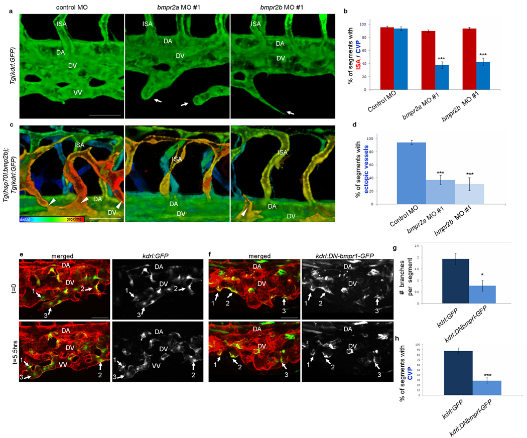
Angiogenesis, the formation of new blood vessels from pre-existing vessels, is critical to most physiological processes and many pathological conditions. During zebrafish development, angiogenesis expands the axial vessels into a complex vascular network that is necessary for efficient oxygen delivery. Although the dorsal aorta and the axial vein are spatially juxtaposed, the initial angiogenic sprouts from these vessels extend in opposite directions, indicating that distinct cues may regulate angiogenesis of the axial vessels. We found that angiogenic sprouts from the dorsal aorta are dependent on vascular endothelial growth factor A (Vegf-A) signalling, and do not respond to bone morphogenetic protein (Bmp) signals. In contrast, sprouts from the axial vein are regulated by Bmp signalling independently of Vegf-A signals, indicating that Bmp is a vein-specific angiogenic cue during early vascular development. Our results support a paradigm whereby different signals regulate distinct programmes of sprouting angiogenesis from the axial vein and dorsal aorta, and indicate that signalling heterogeneity contributes to the complexity of vascular networks.


Figures

Figure 1
Figure 1. The AV forms angiogenic sprouts despite loss of Vegf receptor activity, and expresses Bmp pathway components
(a) Epiflourescent images of 34hpf Tg(kdrl:GFP) control and kdrl/kdr MO injected embryos; insets show higher magnification of the CVP region. Asterisks denote the lack of intersegmental arteries in kdrl/kdr MO injected embryos. Scale bar, 250µm. (b) The percentage of segments that contain an ISA (red bars) or a CVP (blue bars) was quantified in control (n=9) and kdrl/kdr (n=10) MO injected embryos. kdrl/kdr MOs completely blocked the formation of arteries but not veins. Error bars represent mean ± SEM. ***P<0.001 versus control, Student’s t test. (c) Expression pattern of bmp2b, bmpr2a, and bmpr2b in the developing CVP region (black arrowheads) at 32hpf, as detected by in situ hybridization. Cross sections from different 32hpf embryos were taken at the area marked by dashed line. Abbreviations: DA, DA; VV, ventral vein; DV, dorsal vein.
Figure 2
Figure 2. Bmp signaling is necessary and sufficient for sprouting from the AV
(a) Blood vessels in wild-type, Tg(hsp70:noggin3), and Tg(hsp70:bmp2b) embryos in the Tg(kdrl:GFP) transgenic background. The entire vascular network of 42hpf embryos was analyzed using epiflourescent images; dashed boxes represent the trunk and tail areas analyzed below. Z-stacks from the trunk and tail regions were used to make 3-D color projections, where red represents the most proximal (closest to viewer) and blue represents the most distal (farthest from viewer) blood vessels (epiflourescent images and 3-D color projections were taken from different embryos). Scale bar, 50µm. (b)Time lapse imaging of Tg(fli1:nGFP);Tg(kdrl:ras-mCherry) embryos starting at 32hpf. Arrows in panel a and b show sprouts from the AV that fail to make connections in Tg(hsp70:noggin3) embryos. Arrowheads in panel a and b point to ectopic sprouts that branch from the AV in Tg(hsp70:bmp2b) embryos. Scale bar, 20µm. Abbreviations: DA, DA; VV, ventral vein; DV, dorsal vein; NC, notocord; NT, neural tube; ISA, intersegmental artery.
Figure 3
Figure 3. Angiogenesis from the AV requires bmpr2a and bmpr2b and involves endothelial cell autonomous activation of Bmp signaling
(a) Confocal monochrome projections of Tg(kdrl:GFP) embryos injected with a standard control, bmpr2a, or bmpr2b MO. The sprouts from the AV are disrupted with bmpr2a and bmpr2b MO (arrows). (b) The percentage of segments that contain an ISA (red bars) or a CVP (blue bars) was quantified. Total of eight embryos were used for the quantification in each case. bmpr2a or bmpr2b MOs blocked the formation of veins but not arteries. (c) Confocal color depth-code projections of Tg(hsp70l:bmp2b);Tg(kdrl:GFP) heat-shocked embryos injected with a standard control, bmpr2a, or bmpr2b MO. The ectopic sprouts (arrowheads) are reduced in both bmpr2a and bmpr2b morphants. (d) The percentage of segments that contain an ectopic sprout was quantified in control (n=37), bmpr2a #1 (n=27), and bmpr2b #1 (n=15) MO injected embryos. The number of Bmp-induced ectopic sprouts was significantly reduced in both bmpr2a and bmpr2b morphants. (e–f) Time-lapse confocal images of Tg(kdrl:GFP) (e) and Tg(kdrl:DNBmpr1-GFP) (f) mosaic embryos in a Tg(kdrl:ras-mCherry) background. Numbered arrows indicate mosaic endothelial cells. (g) The number of branch points and (h) the percent of segments containing a CVP were quantified in mosaic segment containing GFP or DNBmprI-GFP cells. Total of 29 segments in 7 embryos for Tg(kdrl:GFP) and 34 segments in 11 embryos for Tg(kdrl:DNBmpr1-GFP) were used for quantification (See Methods for detailed quantification method). DNBmprI-GFP-expressing endothelial cells contain fewer branches (g) and fail to form proper CVP connections (h). Scale bar, 50µm. Error bars represent mean ± SEM. **P<0.01 and ***P<0.001 versus control, Student’s t test. Abbreviations: DA, DA; ISA, intersegmental artery; VV, ventral vein; DV, dorsal vein.
Figure 4
Figure 4. Activation of R-Smad and Erk mediates Bmp-induced angiogenesis
(a) Epiflourescent micrographs of Tg(kdrl:GFP) embryos at 38hpf were taken after treatment with DMSO, DMH1 (R-Smad inhibitor), SB203580 (p38 inhibitor), and SL327 (Erk inhibitor). Arrows point to defects in the formation of venous vessels in DMH1- or SL327-treated embryos. (b) The percentage of segments that contain an ISA (red bars) or a CVP (blue bars) was quantified in DMSO (n=14), DMH1 (n=13), SB203580 (n=8), or SL327 (n=10) treated embryos. (c) Confocal depth-code color projections of Tg(hsp70l:bmp2b);Tg(kdrl:GFP) embryos at 46hpf were taken after treatment with small molecule inhibitors. Addition of DMH1 or SL327 to bmp2b over-expressing embryos inhibited Bmp-induced ectopic sprouts. Arrowheads point to ectopic sprouts from the AV. (d) The percentage of segments that contain an ectopic vessel was quantified in DMSO (n=11), DMH1 (n=13), SB203580 (n=4), or SL327 (n=6) treated embryos. (e) The average ectopic vessel length was quantified in DMSO (n=15), DMH1 (n=14), SB203580 (n=16), or SL327 (n= 22) treated embryos. Inhibition of either R-Smad or Erk activation significantly reduced the formation ectopic vessels and the average length of ectopic vessels. Error bars represent mean ± SEM. **P<0.01 and ***P<0.001 versus control, Student’s t test.
Figure 5
Figure 5. Bmp signaling regulates AV angiogenesis independent of Vegf receptor activity
(a) Control and kdrl/kdr MOs were injected into Tg(hsp70:bmp2b); Tg(kdrl:GFP) heat-shocked embryos and shown as 3D color projections. The number of Bmp-induced ectopic sprouts (arrowheads) was not affected by the loss of Kdrl/Kdr activity. Scale bar, 50µm. (b) The percentage of segments that contain ectopic vessels was quantified (n=3 for control, and 6 for kdrl/kdr MO). There was no statistically significant difference between control and kdrl/kdr MOs injected embryos. Error bars represent mean ± SEM. (c) 3-D color projections were taken from the trunk and tail region of 42hpf heat-shocked embryos. Over-expression of bmp2b induced ectopic sprouts in venous endothelial cells (arrowheads), while over-expression of vegfa stimulated ectopic sprouts in arterial endothelial cells in the trunk (arrows). (d) In this model, Bmp signaling is the dominant regulator of AV angiogenesis, while Vegf-A is the main regulator of angiogenesis from the DA. (e) In venous endothelial cells, Bmp2b ligand binds to a Bmpr2a and/or Bmpr2b and Alk2/Alk3 hetero-tetrameric complex, which phosphorylates R-Smad and Erk to promote angiogenesis, while arterial cells utilize the classical Vegf-A signaling cascade to induce angiogenesis. Scale bar, 50µm. Abbreviations: DA, DA; VV, ventral vein; DV, dorsal vein; ISA, intersegmental artery.

References

    1. Isogai S, Lawson N, Torrealday S, Horiguchi M, Weinstein B. Angiogenic network formation in the developing vertebrate trunk. Development. 2003;130:5281–5290. - PubMed
    1. Risau W. Mechanisms of angiogenesis. Nature. 1997;386:671–674. - PubMed
    1. Covassin L, Villefranc J, Kacergis M, Weinstein B, Lawson N. Distinct genetic interactions between multiple Vegf receptors are required for development of different blood vessel types in zebrafish. Proc. Natl. Acad. Sci. U S A. 2006;103:6554–6559. - PMC - PubMed
    1. Herbert S, et al. Arterial-Venous Segregation by Selective Cell Sprouting: An Alternative Mode of Blood Vessel Formation. Science. 2009;326:294–298. - PMC - PubMed
    1. Lee S, et al. Autocrine VEGF signaling is required for vascular homeostasis. Cell. 2007;130:691–703. - PMC - PubMed

Publication types