. 2011 May 15;7(7):469-78.
doi: 10.1038/nchembio.579.

Click-generated triazole ureas as ultrapotent in vivo-active serine hydrolase inhibitors

Affiliations

Click-generated triazole ureas as ultrapotent in vivo-active serine hydrolase inhibitors

Alexander Adibekian et al. Nat Chem Biol. .

Erratum in

  • Nat Chem Biol. 2012 Mar;8(3):318

Abstract

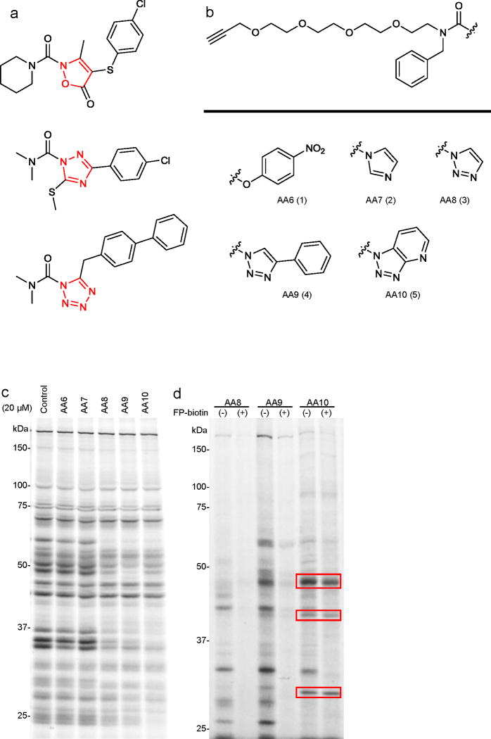
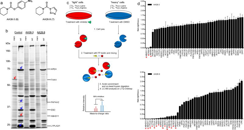
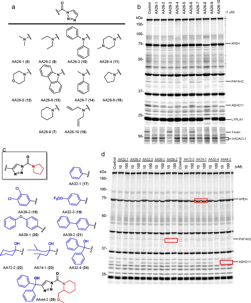
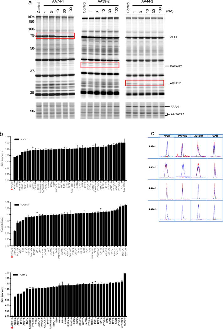
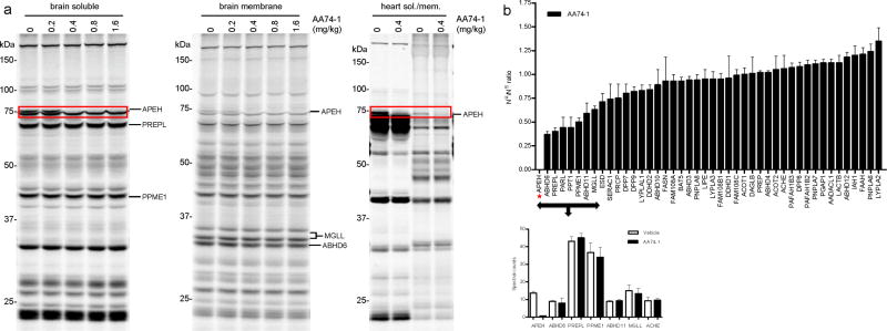
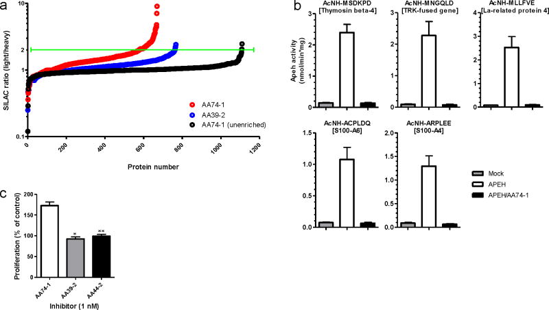
Serine hydrolases are a diverse enzyme class representing ∼1% of all human proteins. The biological functions of most serine hydrolases remain poorly characterized owing to a lack of selective inhibitors to probe their activity in living systems. Here we show that a substantial number of serine hydrolases can be irreversibly inactivated by 1,2,3-triazole ureas, which show negligible cross-reactivity with other protein classes. Rapid lead optimization by click chemistry-enabled synthesis and competitive activity-based profiling identified 1,2,3-triazole ureas that selectively inhibit enzymes from diverse branches of the serine hydrolase class, including peptidases (acyl-peptide hydrolase, or APEH), lipases (platelet-activating factor acetylhydrolase-2, or PAFAH2) and uncharacterized hydrolases (α,β-hydrolase-11, or ABHD11), with exceptional potency in cells (sub-nanomolar) and mice (<1 mg kg(-1)). We show that APEH inhibition leads to accumulation of N-acetylated proteins and promotes proliferation in T cells. These data indicate 1,2,3-triazole ureas are a pharmacologically privileged chemotype for serine hydrolase inhibition, combining broad activity across the serine hydrolase class with tunable selectivity for individual enzymes.


Conflict of interest statement

Competing Financial Interests

The authors declare no competing financial interests.

Figures

Figure 1
Figure 1
Competitive ABPP with clickable N-heterocyclic urea (NHU) activity-based probes AA6-AA10. (a) Structures of previously reported serine hydrolase inhibitors from the NHU class, including the endocannabinoid hydrolase inhibitor tetrazole urea LY2183240 (top) and the HSL inhibitors isoxazolonyl urea (middle) and 1,2,4-triazole urea (bottom). (b) Structures of carbamate- and NHU-alkyne probes with various leaving groups. (c) Competitive ABPP of AA6 - AA10 in the mouse brain membrane proteome. Brain membranes were incubated with 20 μM of AA6 - AA10 or DMSO for 30 min at 37 oC. Proteomes were then labeled with the SH-directed ABPP probe FP-Rh (2 μM, 30 min, 25 °C), separated by SDS-PAGE, and FP-Rh-labeled proteins detected by in-gel fluorescence scanning. This fluorescent gel and all gels in subsequent figures, are shown in grayscale. (d) Profiling the direct targets of AA6 - AA10 (20 μM, 30 min at 37 °C) in brain membranes in the presence or absence of the SH-directed probe FP-biotin (20 μM, 30 min at 37 °C). AA6 - AA10-labeled proteins were detected by reaction with an azide-Rh tag under click chemistry conditions following described protocols,. Targets of AA10 that are not competed by FP-biotin are highlighted with red boxes.
Figure 2
Figure 2
Comparative ABPP of piperidine-based carbamate (AA38-3) and triazole urea (AA26-9) inhibitors. (a) Structures of AA38-3 and AA26-9. (b) Competitive ABPP of AA38-3 and AA26-9 in BW5147-derived murine T-cell hybridoma cells. Cells were cultured with 20 μM inhibitor or DMSO as a control for 4 h, lysed, separated into soluble and analyzed by competitive gel-based ABPP. Blue and red arrows mark SHs that were inhibited by AA26-9 and both AA26-9 and AA38-3, respectively. (c) Schematic representation of a competitive ABPP-SILAC experiment. Isotopically “light” (red) and “heavy” (blue) mouse T-cells are treated with inhibitor and DMSO, respectively, for 4 h. Cells are lysed, proteomes are treated with FP-biotin, and combined in a 1:1 ratio. Biotinylated proteins are enriched, trypsinized, and analyzed by LC-MS/MS. SH activities are quantified by comparing intensities of light and heavy peptide peaks. (d) Identification of SH targets for AA38-3 (top) and AA26-9 (bottom) in mouse T-cells by ABPP-SILAC. Cells were cultured with inhibitor (20 μM) or DMSO as a control for 4 h prior to analysis by competitive ABPP-SILAC. Asterisks mark SHs that were inhibited by > 75%. Bars represent the means ± s.e.m of light/heavy-ratios of identified tryptic peptides in both soluble and membrane proteomes for two independent biological replicates.
Figure 3
Figure 3
Rapid optimization of triazole urea inhibitors by click chemistry-enabled synthesis and competitive ABPP. (a) Structures of ten 1,2,3-triazole ureas (AA26-1-AA26-10) with distinct carbamoyl substituents combined with a uniform, unsubstituted 1,2,3-triazole leaving group. (b) Reactivity profiles for AA26-1-AA26-10 in vitro. Soluble and membrane fractions of mouse T-cells were incubated with inhibitors (1 μM) for 30 min at 37 oC, after which the samples were analyzed by competitive gel-based ABPP. (c) Structures of representative pyrrolidine and piperidine compounds with functionalized 1,2,3-triazole leaving groups. (d) Competitive ABPP results for functionalized 1,2,3-triazole urea inhibitors in mouse T-cells in vitro (treated with inhibitors at the indicated concentrations for 30 min at 37 oC). Inhibitors AA39-2, AA74-1, and AA44-2 each inhibited only a single SH target in the T-cell proteome (highlighted with red boxes).
Figure 4
Figure 4
In vitro and in situ characterization of triazole urea inhibitors AA74-1, AA39-2, and AA44-2 in mouse T-cells. (a) Competitive ABPP results for the three inhibitors in soluble (top) and membrane (bottom) proteomes of T-cells after 30 min treatment at the indicated inhibitor concentrations. Inhibited SHs are highlighted with red boxes. The shown gels are representative of at least three independent biological replicate experiments. (b) ABPP-SILAC analysis of SH activities from inhibitor-treated mouse T-cells (in situ treatment with 3 nM AA74-1, AA39-2 or AA44-2 for 4 h). Asterisks mark the SH target of each compound, each of which was inhibited > 97%. Bars represent the means ± s.e.m of light/heavy-ratios for the multiple peptides observed for each enzyme; data are derived from both soluble and membrane proteomes for two independent biological replicates. (c) Orthogonal selectivity of inhibitors AA74-1, AA39-2, and AA44-2 illustrated by showing heavy and light MS1 peak pairs for representative tryptic peptides from APEH, PAFAH2, ABHD11, and FAAH. Note that unsubstituted inhibitor AA26-9 nonselectively inhibits all four SHs.
Figure 5
Figure 5
Characterization of the activity and selectivity of APEH inhibitor AA74-1 in vivo. (a) Competitive ABPP results for soluble and membrane proteomes from brain and heart tissue of AA74-1-treated mice. Proteomes were prepared from mice injected with AA74-1 (0.2–1.6 mg/kg, i.p.) or vehicle (PEG300) for 4 h and analyzed by competitive gel-based ABPP, n = 3 mice per group. Inhibition of APEH is highlighted with red boxes. (b) ABPP-SILAM analysis of SH activities in brain tissue from mice treated with AA74-1 (0.8 mg/kg, i.p.) or vehicle (PEG300). Asterisk marks the ratio-of-ratio value for APEH, which was inhibited by greater than 97%. Bars represent the means ± s.e.m of ratios-of-ratios for the multiple peptides observed for each enzyme; data are derived from a single experiment.
Figure 6
Figure 6
Proteomic characterization of endogenous APEH substrates using N-terminal labeling and enrichment. (a) Measured SILAC ratios for N-terminally enriched and unenriched peptides from the soluble proteome of mouse T-cells treated in situ with AA74-1 or AA39-2 (1 nM, 6 h). Green line designates the two-fold signal change cut-off used to define candidate APEH substrates in AA74-1-treated cells. (b) In vitro APEH exopeptidase activity assay with synthetic N-acetylated hexapeptides. APEH was recombinantly expressed in HEK-293 cells. Whole cell lysates were pre-treated with DMSO or AA74-1 (3 nM, 30 min), incubated with peptides for 10 h, and release of the N-terminal N-acetylated amino acid was measured by LC-MS. Data are presented as means ± s.d. (n = 3). Mock corresponds to control cells transfected with an empty vector. (c) Stimulation of mouse T-cell proliferation by APEH inhibition. Mouse T-cells were treated in situ with the indicated inhibitors (1 nM) or DMSO for 12 h. Cell proliferation was measured using the colorimetric agent WST-1 (*p < 0.05 for AA74-1-versus AA39-2-treated cells; **p < 0.01 for AA74-1- versus AA44-2-treated cells). Data are presented as means ± s.d. (n = 4).

References

    1. Simon GM, Cravatt BF. Activity-based proteomics of enzyme superfamilies: serine hydrolases as a case study. J Biol Chem. 2010;285:11051–5. - PMC - PubMed
    1. Henness S, Perry CM. Orlistat: a review of its use in the management of obesity. Drugs. 2006;66:1625–56. - PubMed
    1. Thornberry NA, Weber AE. Discovery of JANUVIA (Sitagliptin), a selective dipeptidyl peptidase IV inhibitor for the treatment of type 2 diabetes. Curr Top Med Chem. 2007;7:557–68. - PubMed
    1. Kluge AF, Petter RC. Acylating drugs: redesigning natural covalent inhibitors. Curr Opin Chem Biol. 2010;14:421–7. - PubMed
    1. Birks J, Grimley Evans J, Iakovidou V, Tsolaki M, Holt FE. Rivastigmine for Alzheimer’s disease. Cochrane Database Syst Rev. 2009:CD001191. - PubMed

Publication types

MeSH terms