

Unilateral vogt-koyanagi-harada disease: report of two cases
- PMID: 21572744
- PMCID: PMC3085163
- DOI: 10.4103/0974-9233.75898
Unilateral vogt-koyanagi-harada disease: report of two cases
Abstract
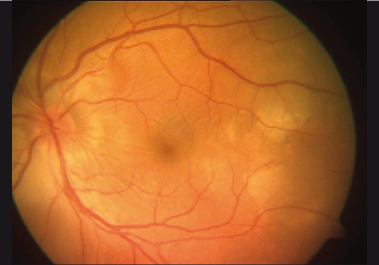
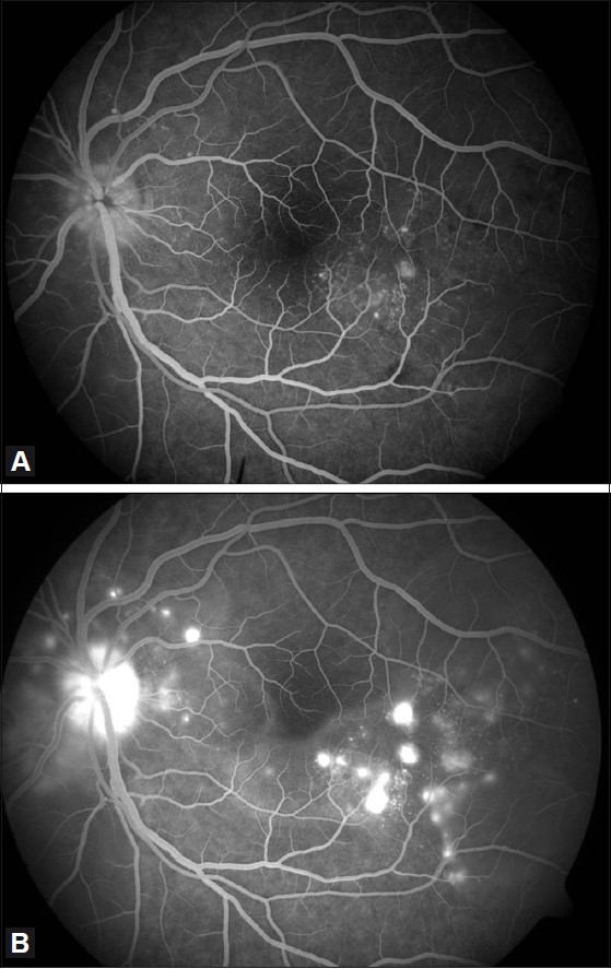
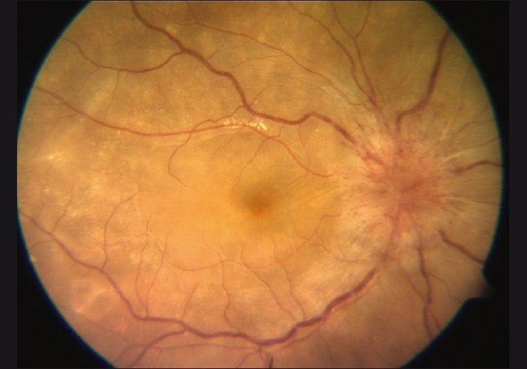
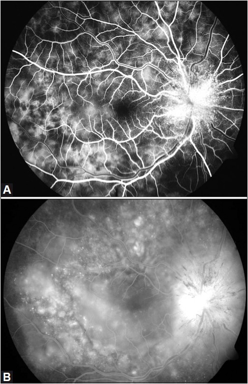
In this retrospective report, we present two cases of unilateral Vogt-Koyanagi-Harada (VKH) disease. These patients were evaluated with clinical, ophthalmological and laboratory examinations. Their response following corticosteroid administration was evaluated. Both patients had the characteristic clinical features of VKH involving only one eye, including disc edema, choroidal striae, multiple sub retinal yellow lesions and exudative retinal detachment. These cases indicate that the clinical and angiographic features were typical of VKH disease despite the unilateral involvement.
Keywords: Choroidal Striae; Disc Edema; Fundus Fluorescein Angiography; Ultrasonography; Vogt-Koyanagi-Harada Disease.
Conflict of interest statement
Figures
References
-
- Usui Y, Goto H, Sakai J, Takeuchi M, Usui M, Rao NA, et al. Presumed Vogt Koyanagi Harada disease with unilateral ocular involvement: Report of three cases. Graefes Arch Clin Exp Opthalmol. 2009;247:1127–32. - PubMed
-
- Forster DJ, Green RL, Rao NA. Unilateral manifestation of Vogt-Koyanagi Harada syndrome in a 7-year old child. Am J Ophthalmol. 1991;111:380–2. - PubMed
-
- Kouda N, Sasaki H, Harada S, Yamada Y, Takahashi N, Sasaki K, et al. Early manifestation of Vogt Koyanagi Harada disease as unilateral posterior scleritis. Jpn J Opthalmol. 2002;46:590–3. - PubMed
LinkOut - more resources
Full Text Sources
