. 2011 May 10;6(5):e18873.
doi: 10.1371/journal.pone.0018873.

Influence of clinical status and parasite load on erythropoiesis and leucopoiesis in dogs naturally infected with leishmania (Leishmania) chagasi

Affiliations

Influence of clinical status and parasite load on erythropoiesis and leucopoiesis in dogs naturally infected with leishmania (Leishmania) chagasi

Raquel Trópia de Abreu et al. PLoS One. .

Abstract

Background: The bone marrow is considered to be an important storage of parasites in Leishmania-infected dogs, although little is known about cellular genesis in this organ during canine visceral leishmaniasis (CVL).

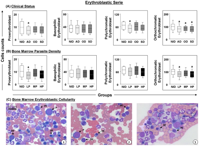
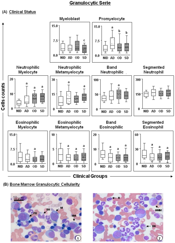
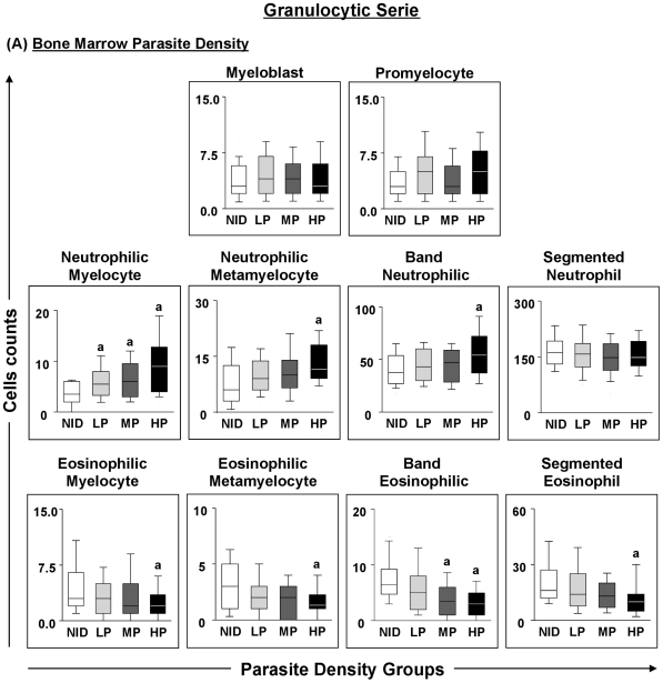
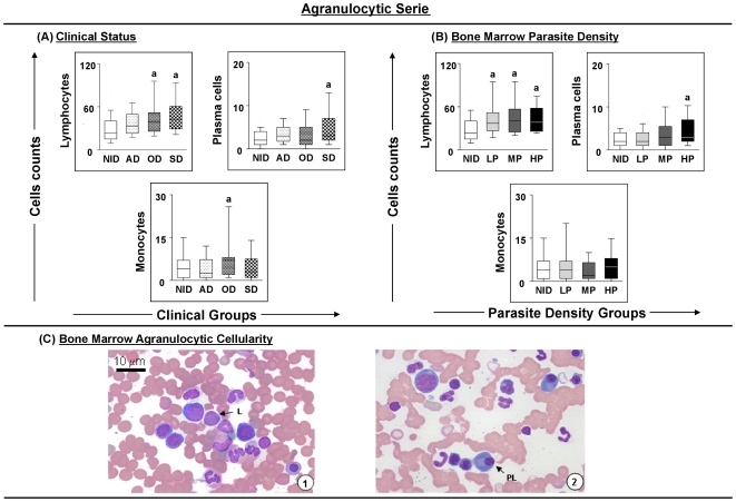
Methodology/principal findings: The aim of the present study was to evaluate changes in erythropoiesis and leucopoiesis in bone marrow aspirates from dogs naturally infected with Leishmania chagasi and presenting different clinical statuses and bone marrow parasite densities. The evolution of CVL from asymptomatic to symptomatic status was accompanied by increasing parasite density in the bone marrow. The impact of bone marrow parasite density on cellularity was similar in dogs at different clinical stages, with animals in the high parasite density group. Erythroid and eosinophilic hypoplasia, proliferation of neutrophilic precursor cells and significant increases in lymphocytes and plasma cell numbers were the major alterations observed. Differential bone marrow cell counts revealed increases in the myeloid:erythroid ratio associated to increased numbers of granulopoietic cells in the different clinical groups compared with non-infected dogs.

Conclusions: Analysis of the data obtained indicated that the assessment of bone marrow constitutes an additional and useful tool by which to elaborate a prognosis for CVL.


Conflict of interest statement

Competing Interests: The authors have declared that no competing interests exist.

Figures

Figure 1
Figure 1. Profiles of the erythroblastic cell series in dogs naturally infected with Leishmania chagasi.
Panel A - animals categorised according to their clinical status into asymptomatic (AD = white boxes with small points), oligosymptomatic (OD = white boxes with large points), symptomatic (SD = white boxes with squares) or non-infected (NID = white boxes) groups. Panel B - animals categorised according to bone marrow parasite density into low (LP = gray boxes), medium (MP = dark gray boxes) and high (HP = black boxes) parasite density and non-infected (NID = white boxes) groups. Absolute cell counts are presented in a box-plot format, the median, maximum and minimum values represents the interquartile range. Significant differences (p<0.05) with respect to the NID group are indicated by the letter ‘a’. Panel C - cellularity of the erythroblastic series observed in the bone marrow of dogs assessed in this study. Slides were stained with Giemsa; bar  = 10 µm. P indicates proerythroblast; BE, basophilic erythroblast; PE, polychromatic erythroblast; OE, orthochromatic erythroblast.
Figure 2
Figure 2. Profiles of the granulocytic cell series in dogs naturally infected with Leishmania chagasi categorised according to clinical status.
Panel A - animals categorised according to their clinical status into asymptomatic (AD = white boxes with small points), oligosymptomatic (OD = white boxes with large points), symptomatic (SD = white boxes with squares) or non-infected (NID = white boxes) groups. Absolute cell counts are presented in a box-plot format, the median, maximum and minimum values represents the interquartile range. Significant differences (p<0.05) with respect to the NID, AD and OD groups are indicated by the letters ‘a’ and ‘b’ and ‘c’ respectively. Panel B - cellularity of the granulocytic series observed in the bone marrow of dogs assessed in this study. Slides were stained with Giemsa; bar  = 10 µm. M indicates myeloblast; PR, promyelocyte; NM, neutrophilic myelocyte; NMM, neutrophilic metamyelocyte; BN band neutrophil; N, neutrophil; EM, eosinophilic myelocyte; E, eosinophil.
Figure 3
Figure 3. Profiles of the granulocytic cell series in dogs naturally infected with Leishmania chagasi categorised according to parasite density.
Panel A - animals categorised according to bone marrow parasite density into low (LP = gray boxes), medium (MP =  dark gray boxes) and high (HP = black boxes) parasite density and non-infected (NID = white boxes) groups. Absolute cell counts are presented in a box-plot format, the median, maximum and minimum values represents the interquartile range. Significant differences (p<0.05) with respect to the NID group are indicated by the letter ‘a’.
Figure 4
Figure 4. Profiles of the agranulocytic cell series in dogs naturally infected with Leishmania chagasi.
Panel A - animals categorised according to their clinical status into asymptomatic (AD = white boxes with small points), oligosymptomatic (OD = white boxes with large points), symptomatic (SD = white boxes with squares) or non-infected (NID = white boxes) groups. Panel B - animals categorised according to bone marrow parasite density into low (LP = gray boxes), medium (MP = dark gray boxes) and high (HP = black boxes) parasite density and non-infected (NID = white boxes) groups. Absolute cell counts are presented in a box-plot format, the median, maximum and minimum values represents the interquartile range. Significant differences (p<0.05) with respect to the NID group are indicated by the letter ‘a’. Panel C - cellularity of the agranulocytic series observed in the bone marrow of dogs assessed in this study. Slides were stained with Giemsa; bar  = 10 µm. L indicates lymphocyte; PC, plasma cell; M, monocyte.

References

    1. Desjeux P. Leishmaniasis: current situation and new perspectives. Comp Immunol Microbiol Infect Dis. 2004;27:305–318. - PubMed
    1. Vieira JB, Coelho GE. [Visceral leishmaniasis or kala-azar: the epidemiological and control aspects]. Rev Soc Bras Med Trop. 1998;31(Suppl 2):85–92. - PubMed
    1. Molina R, Amela C, Nieto J, San-Andres M, Gonzalez F, et al. Infectivity of dogs naturally infected with Leishmania infantum to colonized Phlebotomus perniciosus. Trans R Soc Trop Med Hyg. 1994;88:491–493. - PubMed
    1. Sanchez MA, Diaz NL, Zerpa O, Negron E, Convit J, et al. Organ-specific immunity in canine visceral leishmaniasis: analysis of symptomatic and asymptomatic dogs naturally infected with Leishmania chagasi. Am J Trop Med Hyg. 2004;70:618–624. - PubMed
    1. Reis AB, Martins-Filho OA, Teixeira-Carvalho A, Carvalho MG, Mayrink W, et al. Parasite density and impaired biochemical/hematological status are associated with severe clinical aspects of canine visceral leishmaniasis. Res Vet Sci. 2006;81:68–75. - PubMed

Publication types

LinkOut - more resources